 Movie
Movie Controller

[English] 日本語
 Yorodumi
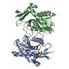
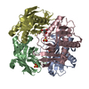
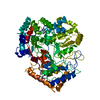
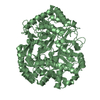
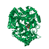
- PDB-2igb: Crystal Structure of PyrR, The Regulator Of The Pyrimidine Biosyn... -
+ Open data

- Basic information
| Entry | Database: PDB / ID: 2igb | ||||||
|---|---|---|---|---|---|---|---|
| Title | Crystal Structure of PyrR, The Regulator Of The Pyrimidine Biosynthetic Operon In Bacillus caldolyticus, UMP-bound form | ||||||
 Components Components | PyrR bifunctional protein | ||||||
 Keywords Keywords | TRANSFERASE / TRANSCRIPTION / TRANSCRIPTION REGULATION / ATTENUATION PROTEIN / RNA-BINDING / PYRIMIDINE BIOSYNTHESIS / PRTASE / URACIL PHOSPHORIBOSYLTRANSFERASE | ||||||
| Function / homology |  Function and homology information Function and homology informationuracil phosphoribosyltransferase / uracil phosphoribosyltransferase activity / DNA-templated transcription termination / RNA binding Similarity search - Function | ||||||
| Biological species |  Bacillus caldolyticus (bacteria) Bacillus caldolyticus (bacteria) | ||||||
| Method |  X-RAY DIFFRACTION / SYNCHROTRON / MOLECULAR REPLACEMENT / Resolution: 1.68 Å X-RAY DIFFRACTION / SYNCHROTRON / MOLECULAR REPLACEMENT / Resolution: 1.68 Å | ||||||
 Authors Authors | Chander, P. / Switzer, R.L. / Smith, J.L. | ||||||
 Citation Citation | Journal: To be Published Title: PyrR, the regulator of the pyrimidine biosynthetic operon in Bacillus caldolyticus Authors: Chander, P. / Switzer, R.L. / Smith, J.L. | ||||||
| History |
|
- Structure visualization
| Structure viewer | Molecule: MolmilJmol/JSmol |
|---|
- Downloads & links
-Download
| PDBx/mmCIF format | 2igb.cif.gz | 90.3 KB | Display | PDBx/mmCIF format |
|---|---|---|---|---|
| PDB format | pdb2igb.ent.gz | 69.1 KB | Display | PDB format |
| PDBx/mmJSON format | 2igb.json.gz | Tree view | PDBx/mmJSON format | |
| Others |  Other downloads Other downloads |
-Validation report
| Arichive directory | https://data.pdbj.org/pub/pdb/validation_reports/ig/2igbftp://data.pdbj.org/pub/pdb/validation_reports/ig/2igb | HTTPS FTP |
|---|
-Related structure data
| Related structure data | 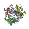 1nonS S: Starting model for refinement |
|---|---|
| Similar structure data |


-Links
 PDBj
PDBj


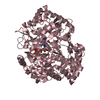
- Assembly
| Deposited unit | 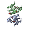
| |||||||||||||||||||||||||||||||||||||||||||||||||||||||||||||||||
|---|---|---|---|---|---|---|---|---|---|---|---|---|---|---|---|---|---|---|---|---|---|---|---|---|---|---|---|---|---|---|---|---|---|---|---|---|---|---|---|---|---|---|---|---|---|---|---|---|---|---|---|---|---|---|---|---|---|---|---|---|---|---|---|---|---|---|
| 1 | 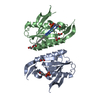
| |||||||||||||||||||||||||||||||||||||||||||||||||||||||||||||||||
| Unit cell |
| |||||||||||||||||||||||||||||||||||||||||||||||||||||||||||||||||
| Noncrystallographic symmetry (NCS) | NCS domain:
NCS domain segments: Ens-ID: 1 / Refine code: 4
|
-Components
| #1: Protein | Mass: 19967.051 Da / Num. of mol.: 2 Source method: isolated from a genetically manipulated source Source: (gene. exp.) Bacillus caldolyticus (bacteria) / Gene: pyrR / Production host: References: UniProt: P41007, uracil phosphoribosyltransferase #2: Chemical |   Mass: 324.181 Da / Num. of mol.: 2 / Source method: obtained synthetically / Formula: C9H13N2O9P Mass: 324.181 Da / Num. of mol.: 2 / Source method: obtained synthetically / Formula: C9H13N2O9P#3: Chemical | ChemComp-EDO /   Mass: 62.068 Da / Num. of mol.: 6 / Source method: obtained synthetically / Formula: C2H6O2 Mass: 62.068 Da / Num. of mol.: 6 / Source method: obtained synthetically / Formula: C2H6O2#4: Water | ChemComp-HOH / |  Mass: 18.015 Da / Num. of mol.: 286 / Source method: isolated from a natural source / Formula: H2O Mass: 18.015 Da / Num. of mol.: 286 / Source method: isolated from a natural source / Formula: H2O |
|---|
-Experimental details
-Experiment
| Experiment | Method: X-RAY DIFFRACTION / Number of used crystals: 1 |
|---|
- Sample preparation
| Crystal | Density Matthews: 2.53 Å3/Da / Density % sol: 51.43 % |
|---|---|
| Crystal grow | Temperature: 298 K / Method: vapor diffusion, hanging drop / pH: 5.5 Details: 0.1M sodium citrate, 0.2M sodium chloride, 1.0M diammonium hydrogen phosphate, pH 5.5, VAPOR DIFFUSION, HANGING DROP, temperature 298K |
-Data collection
| Diffraction source | Source: SYNCHROTRON / Site: APS  / Beamline: 23-ID-B / Wavelength: 0.97 Å / Beamline: 23-ID-B / Wavelength: 0.97 Å |
|---|---|
| Detector | Date: Mar 22, 2006 |
| Radiation | Protocol: SINGLE WAVELENGTH / Monochromatic (M) / Laue (L): M / Scattering type: x-ray |
| Radiation wavelength | Wavelength: 0.97 Å / Relative weight: 1 |
| Reflection | Resolution: 1.68→50 Å / Num. obs: 46630 / Redundancy: 7 % / Biso Wilson estimate: 21.5 Å2 / Rmerge(I) obs: 0.079 / Rsym value: 0.057 |
| Reflection shell | Resolution: 1.68→1.74 Å / Redundancy: 5.5 % / Rmerge(I) obs: 0.93 / Rsym value: 0.62 |
- Processing
| Software |
| ||||||||||||||||||||||||||||||||||||||||||||||||||||||||||||||||||||||||||||||||||||||||||
|---|---|---|---|---|---|---|---|---|---|---|---|---|---|---|---|---|---|---|---|---|---|---|---|---|---|---|---|---|---|---|---|---|---|---|---|---|---|---|---|---|---|---|---|---|---|---|---|---|---|---|---|---|---|---|---|---|---|---|---|---|---|---|---|---|---|---|---|---|---|---|---|---|---|---|---|---|---|---|---|---|---|---|---|---|---|---|---|---|---|---|---|
| Refinement | Method to determine structure: MOLECULAR REPLACEMENT Starting model: PDB entry 1NON Resolution: 1.68→41.31 Å / Cor.coef. Fo:Fc: 0.962 / Cor.coef. Fo:Fc free: 0.946 / SU B: 1.95 / SU ML: 0.066 / Cross valid method: THROUGHOUT / ESU R: 0.1 / ESU R Free: 0.103 / Stereochemistry target values: MAXIMUM LIKELIHOOD / Details: HYDROGENS HAVE BEEN ADDED IN THE RIDING POSITIONS
| ||||||||||||||||||||||||||||||||||||||||||||||||||||||||||||||||||||||||||||||||||||||||||
| Solvent computation | Ion probe radii: 0.8 Å / Shrinkage radii: 0.8 Å / VDW probe radii: 1.4 Å / Solvent model: MASK | ||||||||||||||||||||||||||||||||||||||||||||||||||||||||||||||||||||||||||||||||||||||||||
| Displacement parameters | Biso mean: 23.579 Å2
| ||||||||||||||||||||||||||||||||||||||||||||||||||||||||||||||||||||||||||||||||||||||||||
| Refinement step | Cycle: LAST / Resolution: 1.68→41.31 Å
| ||||||||||||||||||||||||||||||||||||||||||||||||||||||||||||||||||||||||||||||||||||||||||
| Refine LS restraints |
| ||||||||||||||||||||||||||||||||||||||||||||||||||||||||||||||||||||||||||||||||||||||||||
| Refine LS restraints NCS | Dom-ID: 1 / Auth asym-ID: A / Ens-ID: 1 / Number: 932 / Refine-ID: X-RAY DIFFRACTION
| ||||||||||||||||||||||||||||||||||||||||||||||||||||||||||||||||||||||||||||||||||||||||||
| LS refinement shell | Resolution: 1.68→1.73 Å / Total num. of bins used: 20
|
