 Movie
Movie Controller

[English] 日本語
 Yorodumi
- PDB-1non: PyrR, the regulator of the pyrimidine biosynthetic operon in Baci... -
+ Open data

- Basic information
| Entry | Database: PDB / ID: 1non | ||||||
|---|---|---|---|---|---|---|---|
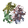
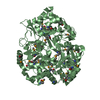
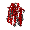
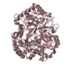
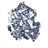
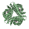
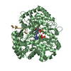
| Title | PyrR, the regulator of the pyrimidine biosynthetic operon in Bacillus caldolyticus | ||||||
 Components Components | PyrR bifunctional protein | ||||||
 Keywords Keywords | TRANSCRIPTION / TRANSFERASE / TRANSCRIPTION REGULATION / ATTENUATION PROTEIN / RNA-BINDING PROTEIN / PYRIMIDINE BIOSYNTHESIS / PRTASE / URACIL PHOSPHORIBOSYLTRANSFERASE / BIFUNCTIONAL ENZYME | ||||||
| Function / homology |  Function and homology information Function and homology informationuracil phosphoribosyltransferase / uracil phosphoribosyltransferase activity / DNA-templated transcription termination / RNA binding Similarity search - Function | ||||||
| Biological species |  Bacillus caldolyticus (bacteria) Bacillus caldolyticus (bacteria) | ||||||
| Method |  X-RAY DIFFRACTION / MOLECULAR REPLACEMENT / Resolution: 2.4 Å X-RAY DIFFRACTION / MOLECULAR REPLACEMENT / Resolution: 2.4 Å | ||||||
 Authors Authors | Switzer, R.L. / Chander, P. / Smith, J.L. / Halbig, K.M. / Miller, J.K. / Bonner, H.K. / Grabner, G.K. | ||||||
 Citation Citation | Journal: J.Bacteriol. / Year: 2005 Title: Structure of the Nucleotide Complex of PyrR, the pyr Attenuation Protein from Bacillus caldolyticus, Suggests Dual Regulation by Pyrimidine and Purine Nucleotides Authors: Chander, P. / Halbig, K.M. / Miller, J.K. / Fields, C.J. / Bonner, H.K. / Grabner, G.K. / Switzer, R.L. / Smith, J.L. | ||||||
| History |
|
- Structure visualization
| Structure viewer | Molecule: MolmilJmol/JSmol |
|---|
- Downloads & links
-Download
| PDBx/mmCIF format | 1non.cif.gz | 140.5 KB | Display | PDBx/mmCIF format |
|---|---|---|---|---|
| PDB format | pdb1non.ent.gz | 112 KB | Display | PDB format |
| PDBx/mmJSON format | 1non.json.gz | Tree view | PDBx/mmJSON format | |
| Others |  Other downloads Other downloads |
-Validation report
| Arichive directory | https://data.pdbj.org/pub/pdb/validation_reports/no/1nonftp://data.pdbj.org/pub/pdb/validation_reports/no/1non | HTTPS FTP |
|---|
-Related structure data
| Related structure data |  1xz8C 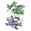 1xznC 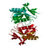 1a3cS S: Starting model for refinement C: citing same article ( |
|---|---|
| Similar structure data |


-Links
 PDBj
PDBj
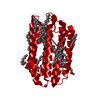
- Assembly
| Deposited unit | 
| ||||||||||
|---|---|---|---|---|---|---|---|---|---|---|---|
| 1 |
| ||||||||||
| Unit cell |
| ||||||||||
| Details | biological aassembley is a tetramer; generated from the monomer by the operation X,Y,Z; -X,Y,-Z; 1/2+X,1/2+Y,Z; 1/2-X,1/2+Y,-Z |
-Components
| #1: Protein | Mass: 19967.051 Da / Num. of mol.: 4 Source method: isolated from a genetically manipulated source Details: includes Pyrimidine operon regulatory protein and Uracil phosphoribosyltransferase Source: (gene. exp.) Bacillus caldolyticus (bacteria) / Gene: pyrr / Plasmid: pSHCO2 / Species (production host): Escherichia coli / Production host: References: UniProt: P41007, uracil phosphoribosyltransferase #2: Water | ChemComp-HOH / |  Mass: 18.015 Da / Num. of mol.: 358 / Source method: isolated from a natural source / Formula: H2O Mass: 18.015 Da / Num. of mol.: 358 / Source method: isolated from a natural source / Formula: H2O |
|---|
-Experimental details
-Experiment
| Experiment | Method: X-RAY DIFFRACTION / Number of used crystals: 1 |
|---|
- Sample preparation
| Crystal | Density Matthews: 2.28 Å3/Da / Density % sol: 45.6 % |
|---|---|
| Crystal grow | Temperature: 298 K / Method: vapor diffusion, hanging drop / pH: 7.4 Details: PEG 400, ammonium chloride, magnesium chloride, cacodylate, pH 7.4, VAPOR DIFFUSION, HANGING DROP, temperature 298K |
-Data collection
| Diffraction | Mean temperature: 120 K |
|---|---|
| Diffraction source | Source: ROTATING ANODE / Type: RIGAKU / Wavelength: 1.5418 Å |
| Detector | Type: RIGAKU RAXIS IV / Detector: IMAGE PLATE / Date: Jan 30, 2002 |
| Radiation | Monochromator: graphite / Protocol: SINGLE WAVELENGTH / Monochromatic (M) / Laue (L): M / Scattering type: x-ray |
| Radiation wavelength | Wavelength: 1.5418 Å / Relative weight: 1 |
| Reflection | Resolution: 2.4→50 Å / Num. all: 26925 / Num. obs: 25768 / % possible obs: 98.2 % / Observed criterion σ(I): -3 / Redundancy: 4 % / Biso Wilson estimate: 52.3 Å2 / Rmerge(I) obs: 0.035 / Rsym value: 0.211 / Net I/σ(I): 19.1 |
| Reflection shell | Resolution: 2.4→2.5 Å / Redundancy: 1 % / Rmerge(I) obs: 0.19 / Mean I/σ(I) obs: 19.1 / Num. unique all: 2376 / Rsym value: 0.211 / % possible all: 92 |
- Processing
| Software |
| |||||||||||||||||||||||||
|---|---|---|---|---|---|---|---|---|---|---|---|---|---|---|---|---|---|---|---|---|---|---|---|---|---|---|
| Refinement | Method to determine structure: MOLECULAR REPLACEMENT Starting model: PDB ENTRY 1A3C Resolution: 2.4→50 Å / Cross valid method: THROUGHOUT / σ(F): 2.4 / Stereochemistry target values: Engh & Huber
| |||||||||||||||||||||||||
| Refinement step | Cycle: LAST / Resolution: 2.4→50 Å
| |||||||||||||||||||||||||
| Refine LS restraints |
|
