 Movie
Movie Controller

[English] 日本語
 Yorodumi
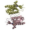
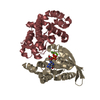
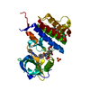
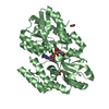
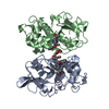
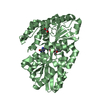
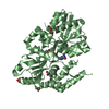
- PDB-1xzn: PYRR, THE REGULATOR OF THE PYRIMIDINE BIOSYNTHETIC OPERON IN BACI... -
+ Open data

- Basic information
| Entry | Database: PDB / ID: 1xzn | ||||||
|---|---|---|---|---|---|---|---|
| Title | PYRR, THE REGULATOR OF THE PYRIMIDINE BIOSYNTHETIC OPERON IN BACILLUS CALDOLYTICUS, sulfate-bound form | ||||||
 Components Components | PyrR bifunctional protein | ||||||
 Keywords Keywords | TRANSCRIPTION / TRANSFERASE / TRANSCRIPTION REGULATION / ATTENUATION PROTEIN / RNA-BINDING / PYRIMIDINE BIOSYNTHESIS / PRTASE / URACIL PHOSPHORIBOSYLTRANSFERASE / BIFUNCTIONAL ENZYME | ||||||
| Function / homology |  Function and homology information Function and homology informationuracil phosphoribosyltransferase / uracil phosphoribosyltransferase activity / DNA-templated transcription termination / RNA binding Similarity search - Function | ||||||
| Biological species |  Bacillus caldolyticus (bacteria) Bacillus caldolyticus (bacteria) | ||||||
| Method |  X-RAY DIFFRACTION / SYNCHROTRON / MOLECULAR REPLACEMENT / Resolution: 2.27 Å X-RAY DIFFRACTION / SYNCHROTRON / MOLECULAR REPLACEMENT / Resolution: 2.27 Å | ||||||
 Authors Authors | Chander, P. / Halbig, K.M. / Miller, J.K. / Fields, C.J. / Bonner, H.K. / Grabner, G.K. / Switzer, R.L. / Smith, J.L. | ||||||
 Citation Citation | Journal: J.Bacteriol. / Year: 2005 Title: Structure of the Nucleotide Complex of PyrR, the pyr Attenuation Protein from Bacillus caldolyticus, Suggests Dual Regulation by Pyrimidine and Purine Nucleotides. Authors: Chander, P. / Halbig, K.M. / Miller, J.K. / Fields, C.J. / Bonner, H.K. / Grabner, G.K. / Switzer, R.L. / Smith, J.L. | ||||||
| History |
| ||||||
| Remark 300 | BIOMOLECULE: 1 THIS ENTRY CONTAINS THE CRYSTALLOGRAPHIC ASYMMETRIC UNIT WHICH CONSISTS OF 2 CHAIN(S) ...BIOMOLECULE: 1 THIS ENTRY CONTAINS THE CRYSTALLOGRAPHIC ASYMMETRIC UNIT WHICH CONSISTS OF 2 CHAIN(S). THE AUTHORS ARE NOT SURE ABOUT WHAT THE BIOLOGICAL MOLECULE FOR THE PROTEIN IS. |
- Structure visualization
| Structure viewer | Molecule: MolmilJmol/JSmol |
|---|
- Downloads & links
-Download
| PDBx/mmCIF format | 1xzn.cif.gz | 76.9 KB | Display | PDBx/mmCIF format |
|---|---|---|---|---|
| PDB format | pdb1xzn.ent.gz | 57.8 KB | Display | PDB format |
| PDBx/mmJSON format | 1xzn.json.gz | Tree view | PDBx/mmJSON format | |
| Others |  Other downloads Other downloads |
-Validation report
| Arichive directory | https://data.pdbj.org/pub/pdb/validation_reports/xz/1xznftp://data.pdbj.org/pub/pdb/validation_reports/xz/1xzn | HTTPS FTP |
|---|
-Related structure data
| Related structure data | 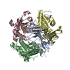 1nonSC  1xz8C S: Starting model for refinement C: citing same article ( |
|---|---|
| Similar structure data |


-Links
 PDBj
PDBj
- Assembly
| Deposited unit | 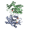
| ||||||||
|---|---|---|---|---|---|---|---|---|---|
| 1 |
| ||||||||
| Unit cell |
|
-Components
| #1: Protein | Mass: 19967.051 Da / Num. of mol.: 2 Source method: isolated from a genetically manipulated source Source: (gene. exp.) Bacillus caldolyticus (bacteria) / Plasmid: PSHCO2 / Species (production host): Escherichia coli / Production host: References: UniProt: P41007, uracil phosphoribosyltransferase #2: Chemical |   Mass: 24.305 Da / Num. of mol.: 2 / Source method: obtained synthetically / Formula: Mg Mass: 24.305 Da / Num. of mol.: 2 / Source method: obtained synthetically / Formula: Mg#3: Chemical |   Mass: 96.063 Da / Num. of mol.: 3 / Source method: obtained synthetically / Formula: SO4 Mass: 96.063 Da / Num. of mol.: 3 / Source method: obtained synthetically / Formula: SO4#4: Water | ChemComp-HOH / |  Mass: 18.015 Da / Num. of mol.: 83 / Source method: isolated from a natural source / Formula: H2O Mass: 18.015 Da / Num. of mol.: 83 / Source method: isolated from a natural source / Formula: H2O |
|---|
-Experimental details
-Experiment
| Experiment | Method: X-RAY DIFFRACTION / Number of used crystals: 1 |
|---|
- Sample preparation
| Crystal | Density Matthews: 2 Å3/Da / Density % sol: 38.5 % |
|---|---|
| Crystal grow | Temperature: 298 K / Method: vapor diffusion, hanging drop / pH: 7.4 Details: 100mM Na cacodylate, 100mM Amm sulfate, 2mM Mg sulfate, 10% Pek 4000, pH 7.4, VAPOR DIFFUSION, HANGING DROP, temperature 298.0K |
-Data collection
| Diffraction source | Source: SYNCHROTRON / Site: APS  / Beamline: 14-ID-B / Beamline: 14-ID-B |
|---|---|
| Radiation | Protocol: SINGLE WAVELENGTH / Monochromatic (M) / Laue (L): M / Scattering type: x-ray |
| Radiation wavelength | Relative weight: 1 |
| Reflection | Resolution: 2.27→37.81 Å / Num. all: 16954 / Num. obs: 16124 / % possible obs: 95.1 % / Observed criterion σ(F): 0 / Observed criterion σ(I): 0 / Redundancy: 10 % / Rmerge(I) obs: 0.094 / Rsym value: 0.312 / Net I/σ(I): 20.6 |
- Processing
| Software |
| ||||||||||||||||||||
|---|---|---|---|---|---|---|---|---|---|---|---|---|---|---|---|---|---|---|---|---|---|
| Refinement | Method to determine structure: MOLECULAR REPLACEMENT Starting model: PDB entry 1NON Resolution: 2.27→50 Å / Stereochemistry target values: Engh & Huber
| ||||||||||||||||||||
| Refinement step | Cycle: LAST / Resolution: 2.27→50 Å
| ||||||||||||||||||||
| Refine LS restraints |
|
