 Movie
Movie Controller


+ Open data

- Basic information
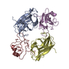
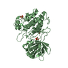
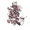
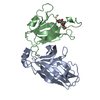
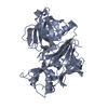
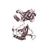
| Entry | Database: PDB / ID: 3gxu | ||||||
|---|---|---|---|---|---|---|---|
| Title | Crystal structure of Eph receptor and ephrin complex | ||||||
 Components Components |
| ||||||
 Keywords Keywords | TRANSFERASE / Complex structure / Eph / ephrin / ATP-binding / Glycoprotein / Kinase / Membrane / Nucleotide-binding / Phosphoprotein / Receptor / Transmembrane / Tyrosine-protein kinase / Developmental protein / Differentiation / Disulfide bond / Host-virus interaction / Neurogenesis | ||||||
| Function / homology |  Function and homology information Function and homology informationDH domain binding / neuron projection fasciculation / : / corticospinal tract morphogenesis / regulation of astrocyte differentiation / neuron projection guidance / venous blood vessel morphogenesis / nephric duct morphogenesis / fasciculation of motor neuron axon / positive regulation of aorta morphogenesis ...DH domain binding / neuron projection fasciculation / : / corticospinal tract morphogenesis / regulation of astrocyte differentiation / neuron projection guidance / venous blood vessel morphogenesis / nephric duct morphogenesis / fasciculation of motor neuron axon / positive regulation of aorta morphogenesis / fasciculation of sensory neuron axon / synapse pruning / negative regulation of cellular response to hypoxia / transmembrane-ephrin receptor activity / positive regulation of leukocyte adhesion to arterial endothelial cell / glial cell migration / presynapse assembly / regulation of modification of synaptic structure / PH domain binding / positive regulation of amyloid precursor protein catabolic process / GPI-linked ephrin receptor activity / regulation of synapse pruning / lymph vessel development / regulation of chemotaxis / ephrin receptor activity / negative regulation of axon regeneration / positive regulation of cardiac muscle cell differentiation / adherens junction organization / regulation of dendritic spine morphogenesis / negative regulation of cell adhesion / cell migration involved in sprouting angiogenesis / regulation of GTPase activity / positive regulation of dendrite morphogenesis / motor neuron axon guidance / EPH-Ephrin signaling / blood vessel morphogenesis / innervation / Ephrin signaling / adult walking behavior / positive regulation of amyloid-beta formation / negative regulation of epithelial to mesenchymal transition / EPHA-mediated growth cone collapse / regulation of postsynaptic neurotransmitter receptor internalization / regulation of axonogenesis / Somitogenesis / negative regulation of long-term synaptic potentiation / positive regulation of intracellular signal transduction / keratinocyte proliferation / cochlea development / EPH-ephrin mediated repulsion of cells / negative regulation of keratinocyte proliferation / ephrin receptor signaling pathway / anatomical structure morphogenesis / axonal growth cone / regulation of postsynaptic membrane neurotransmitter receptor levels / ephrin receptor binding / T cell costimulation / EPHB-mediated forward signaling / positive regulation of cell adhesion / axon terminus / protein tyrosine kinase binding / negative regulation of cell migration / axon guidance / animal organ morphogenesis / dendritic shaft / filopodium / adherens junction / neuromuscular junction / receptor protein-tyrosine kinase / negative regulation of ERK1 and ERK2 cascade / positive regulation of JNK cascade / postsynaptic density membrane / Schaffer collateral - CA1 synapse / cellular response to amyloid-beta / kinase activity / negative regulation of neuron projection development / amyloid-beta binding / virus receptor activity / cellular response to lipopolysaccharide / protein tyrosine kinase activity / angiogenesis / presynaptic membrane / early endosome membrane / dendritic spine / negative regulation of neuron apoptotic process / perikaryon / mitochondrial outer membrane / cell adhesion / negative regulation of translation / protein stabilization / positive regulation of cell migration / receptor ligand activity / axon / focal adhesion / positive regulation of cell population proliferation / dendrite / glutamatergic synapse / cell surface / ATP binding / identical protein binding Similarity search - Function | ||||||
| Biological species |  Homo sapiens (human) Homo sapiens (human) | ||||||
| Method |  X-RAY DIFFRACTION / MOLECULAR REPLACEMENT / Resolution: 2.5 Å X-RAY DIFFRACTION / MOLECULAR REPLACEMENT / Resolution: 2.5 Å | ||||||
 Authors Authors | Qin, H.N. / Song, J.X. | ||||||
 Citation Citation | Journal: J.Biol.Chem. / Year: 2009 Title: Structural characterization of the EphA4-ephrin-B2 complex reveals new features enabling Eph-ephrin binding promiscuity Authors: Qin, H. / Noberini, R. / Huan, X. / Shi, J. / Pasquale, E.B. / Song, J. #1: Journal: J.Biol.Chem. / Year: 2008Title: Crystal structure and NMR binding reveal that two small molecule antagonists target the high affinity ephrin-binding channel of the EphA4 receptor Authors: Qin, H. / Shi, J. / Noberini, R. / Pasquale, E.B. / Song, J. #2: Journal: Nature / Year: 2001Title: Crystal structure of an Eph receptor-ephrin complex Authors: Himanen, J.P. / Rajashankar, K.R. / Lackmann, M. / Cowan, C.A. / Henkemeyer, M. / Nikolov, D.B. | ||||||
| History |
|
- Structure visualization
| Structure viewer | Molecule: MolmilJmol/JSmol |
|---|
- Downloads & links
-Download
| PDBx/mmCIF format | 3gxu.cif.gz | 83 KB | Display | PDBx/mmCIF format |
|---|---|---|---|---|
| PDB format | pdb3gxu.ent.gz | 61.7 KB | Display | PDB format |
| PDBx/mmJSON format | 3gxu.json.gz | Tree view | PDBx/mmJSON format | |
| Others |  Other downloads Other downloads |
-Validation report
| Arichive directory | https://data.pdbj.org/pub/pdb/validation_reports/gx/3gxuftp://data.pdbj.org/pub/pdb/validation_reports/gx/3gxu | HTTPS FTP |
|---|
-Related structure data


-Links
 PDBj
PDBj


- Assembly
| Deposited unit | 
| ||||||||
|---|---|---|---|---|---|---|---|---|---|
| 1 |
| ||||||||
| Unit cell |
|
-Components
| #1: Protein | Mass: 20206.844 Da / Num. of mol.: 1 / Fragment: UNP residues 29-203 Source method: isolated from a genetically manipulated source Source: (gene. exp.) Homo sapiens (human) / Gene: EPHA4, HEK8, SEK, TYRO1 / Plasmid: PET32a / Production host:  References: UniProt: P54764, receptor protein-tyrosine kinase |
|---|---|
| #2: Protein | Mass: 16261.525 Da / Num. of mol.: 1 / Fragment: UNP residues 27-169 Source method: isolated from a genetically manipulated source Source: (gene. exp.) Homo sapiens (human) / Gene: EFNB2, EPLG5, HTKL, LERK5 / Plasmid: PET32a / Production host: |
| #3: Water | ChemComp-HOH /  Mass: 18.015 Da / Num. of mol.: 337 / Source method: isolated from a natural source / Formula: H2O Mass: 18.015 Da / Num. of mol.: 337 / Source method: isolated from a natural source / Formula: H2O |
| Has protein modification | Y |
-Experimental details
-Experiment
| Experiment | Method: X-RAY DIFFRACTION / Number of used crystals: 1 |
|---|
- Sample preparation
| Crystal | Density Matthews: 2.21 Å3/Da / Density % sol: 44.22 % |
|---|---|
| Crystal grow | Temperature: 298 K / Method: vapor diffusion, hanging drop / pH: 7.5 Details: 23.5% PEG4000, 0.1M Tris, 0.2M MgCl2, pH7.5, VAPOR DIFFUSION, HANGING DROP, temperature 298K |
-Data collection
| Diffraction | Mean temperature: 100 K |
|---|---|
| Diffraction source | Source: ROTATING ANODE / Type: BRUKER AXS MICROSTAR-H |
| Radiation | Protocol: SINGLE WAVELENGTH / Monochromatic (M) / Laue (L): M / Scattering type: x-ray |
| Radiation wavelength | Relative weight: 1 |
| Reflection | Resolution: 2.5→50 Å / Num. all: 11071 / Num. obs: 10938 |
- Processing
| Software |
| ||||||||||||||||||||
|---|---|---|---|---|---|---|---|---|---|---|---|---|---|---|---|---|---|---|---|---|---|
| Refinement | Method to determine structure: MOLECULAR REPLACEMENT Starting model: PDB ENTRY 3CKH, 1KGY Resolution: 2.5→25 Å / σ(F): 3 / σ(I): 3 / Stereochemistry target values: Engh & Huber
| ||||||||||||||||||||
| Refinement step | Cycle: LAST / Resolution: 2.5→25 Å
|
