 Movie
Movie Controller

[English] 日本語
 Yorodumi
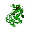
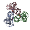
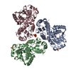
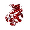
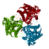
- PDB-2i6u: Crystal Structure of Ornithine Carbamoyltransferase complexed wit... -
+ Open data

- Basic information
| Entry | Database: PDB / ID: 2i6u | ||||||
|---|---|---|---|---|---|---|---|
| Title | Crystal Structure of Ornithine Carbamoyltransferase complexed with Carbamoyl Phosphate and L-Norvaline from Mycobacterium tuberculosis (Rv1656) at 2.2 A | ||||||
 Components Components | Ornithine carbamoyltransferase | ||||||
 Keywords Keywords | TRANSFERASE / Mycobacterium tuberculosis / ornithine carbamyoltransferase / Carbamoyl phosphate / L-Norvaline / Structural Genomics / PSI / Protein Structure Initiative / TB Structural Genomics Consortium / TBSGC | ||||||
| Function / homology |  Function and homology information Function and homology informationornithine carbamoyltransferase / ornithine carbamoyltransferase activity / L-citrulline biosynthetic process / : / L-arginine biosynthetic process / amino acid binding / cytoplasm Similarity search - Function | ||||||
| Biological species |   Mycobacterium tuberculosis (bacteria) Mycobacterium tuberculosis (bacteria) | ||||||
| Method |  X-RAY DIFFRACTION / SYNCHROTRON / MOLECULAR REPLACEMENT / Resolution: 2.2 Å X-RAY DIFFRACTION / SYNCHROTRON / MOLECULAR REPLACEMENT / Resolution: 2.2 Å | ||||||
 Authors Authors | Sankaranarayanan, R. / Moradian, F. / Cherney, L.T. / Garen, C. / Cherney, M.M. / James, M.N.G. / TB Structural Genomics Consortium (TBSGC) | ||||||
 Citation Citation | Journal: J.Mol.Biol. / Year: 2008 Title: The crystal structures of ornithine carbamoyltransferase from Mycobacterium tuberculosis and its ternary complex with carbamoyl phosphate and L-norvaline reveal the enzyme's catalytic mechanism Authors: Sankaranarayanan, R. / Cherney, M.M. / Cherney, L.T. / Garen, C.R. / Moradian, F. / James, M.N. | ||||||
| History |
| ||||||
| Remark 999 | SEQUENCE VAL1 IS IN THE PROTEIN SEQUENCE LISTED AT THE TB CONSORTIUM WEBSITE UNDER THE ACCESSION CODE RV1656. |
- Structure visualization
| Structure viewer | Molecule: MolmilJmol/JSmol |
|---|
- Downloads & links
-Download
| PDBx/mmCIF format | 2i6u.cif.gz | 195.3 KB | Display | PDBx/mmCIF format |
|---|---|---|---|---|
| PDB format | pdb2i6u.ent.gz | 155.4 KB | Display | PDB format |
| PDBx/mmJSON format | 2i6u.json.gz | Tree view | PDBx/mmJSON format | |
| Others |  Other downloads Other downloads |
-Validation report
| Arichive directory | https://data.pdbj.org/pub/pdb/validation_reports/i6/2i6uftp://data.pdbj.org/pub/pdb/validation_reports/i6/2i6u | HTTPS FTP |
|---|
-Related structure data
| Related structure data |  2p2gC  1fb5S S: Starting model for refinement C: citing same article ( |
|---|---|
| Similar structure data | |
| Other databases |

-Links
 PDBj
PDBj
- Assembly
| Deposited unit | 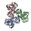
| ||||||||
|---|---|---|---|---|---|---|---|---|---|
| 1 |
| ||||||||
| Unit cell |
| ||||||||
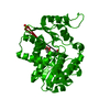
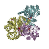
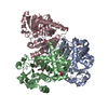
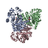
| Details | Assymetric Unit of the crystal contains Trimer which is the Biological entity |
-Components
| #1: Protein | Mass: 33064.195 Da / Num. of mol.: 3 Source method: isolated from a genetically manipulated source Source: (gene. exp.) Mycobacterium tuberculosis (bacteria) / Gene: argF, Rv1656, MT1694, MTCY06H11.21 / Plasmid: PDEST17 / Production host: References: UniProt: P0A5M8, UniProt: P9WIT9*PLUS, ornithine carbamoyltransferase #2: Chemical | ChemComp-SO4 /   Mass: 96.063 Da / Num. of mol.: 5 / Source method: obtained synthetically / Formula: SO4 Mass: 96.063 Da / Num. of mol.: 5 / Source method: obtained synthetically / Formula: SO4#3: Chemical | 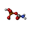  Mass: 141.020 Da / Num. of mol.: 3 / Source method: obtained synthetically / Formula: CH4NO5P Mass: 141.020 Da / Num. of mol.: 3 / Source method: obtained synthetically / Formula: CH4NO5P#4: Chemical | 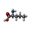  Type: L-peptide linking / Mass: 117.146 Da / Num. of mol.: 3 / Source method: obtained synthetically / Formula: C5H11NO2 Type: L-peptide linking / Mass: 117.146 Da / Num. of mol.: 3 / Source method: obtained synthetically / Formula: C5H11NO2#5: Water | ChemComp-HOH / |  Mass: 18.015 Da / Num. of mol.: 572 / Source method: isolated from a natural source / Formula: H2O Mass: 18.015 Da / Num. of mol.: 572 / Source method: isolated from a natural source / Formula: H2O |
|---|
-Experimental details
-Experiment
| Experiment | Method: X-RAY DIFFRACTION / Number of used crystals: 1 |
|---|
- Sample preparation
| Crystal | Density Matthews: 3.31 Å3/Da / Density % sol: 62.84 % |
|---|---|
| Crystal grow | Temperature: 273 K / Method: vapor diffusion, hanging drop / pH: 6.5 Details: 1.6M Magnesium Sulfate, 0.1M Bis-Tris, 8% PEG 400, 3% ethanol, 3mM L-Norvaline, 3mM Carbamoyl phosphate, pH 6.5, VAPOR DIFFUSION, HANGING DROP, temperature 273K |
-Data collection
| Diffraction | Mean temperature: 100 K |
|---|---|
| Diffraction source | Source: SYNCHROTRON / Site: SSRL  / Beamline: BL9-1 / Wavelength: 0.979462 Å / Beamline: BL9-1 / Wavelength: 0.979462 Å |
| Detector | Type: ADSC QUANTUM 315 / Detector: CCD / Date: Jul 13, 2006 Details: Vertical focusing mirror; single crystal Si(311) bent monochromator (horizontal focusing) |
| Radiation | Monochromator: Side-scattering cuberoot I-beam bent single crystal; asymetric cut 12.2 degs. Protocol: SINGLE WAVELENGTH / Monochromatic (M) / Laue (L): M / Scattering type: x-ray |
| Radiation wavelength | Wavelength: 0.979462 Å / Relative weight: 1 |
| Reflection | Resolution: 2.2→49.568 Å / Num. all: 69827 / Num. obs: 51538 / % possible obs: 73.8 % / Observed criterion σ(F): 0 / Observed criterion σ(I): 0 / Redundancy: 5.5 % / Biso Wilson estimate: 24.1 Å2 / Rmerge(I) obs: 0.082 / Net I/σ(I): 16.4 |
| Reflection shell | Resolution: 2.2→2.28 Å / Redundancy: 3.1 % / Rmerge(I) obs: 0.307 / Mean I/σ(I) obs: 3.3 / Num. unique all: 2981 / % possible all: 43.9 |
- Processing
| Software |
| ||||||||||||||||||||||||||||||||||||||||||||||||||||||||||||||||||||||||||||||||||||||||||
|---|---|---|---|---|---|---|---|---|---|---|---|---|---|---|---|---|---|---|---|---|---|---|---|---|---|---|---|---|---|---|---|---|---|---|---|---|---|---|---|---|---|---|---|---|---|---|---|---|---|---|---|---|---|---|---|---|---|---|---|---|---|---|---|---|---|---|---|---|---|---|---|---|---|---|---|---|---|---|---|---|---|---|---|---|---|---|---|---|---|---|---|
| Refinement | Method to determine structure: MOLECULAR REPLACEMENT Starting model: PDB ENTRY 1FB5 Resolution: 2.2→49.568 Å / Cor.coef. Fo:Fc: 0.95 / Cor.coef. Fo:Fc free: 0.903 / SU B: 4.93 / SU ML: 0.125 / Isotropic thermal model: Isotropic / Cross valid method: THROUGHOUT / σ(F): 0 / σ(I): 0 / ESU R: 0.28 / ESU R Free: 0.233 / Stereochemistry target values: MAXIMUM LIKELIHOOD / Details: HYDROGENS HAVE BEEN ADDED IN THE RIDING POSITIONS
| ||||||||||||||||||||||||||||||||||||||||||||||||||||||||||||||||||||||||||||||||||||||||||
| Solvent computation | Ion probe radii: 0.8 Å / Shrinkage radii: 0.8 Å / VDW probe radii: 1.2 Å / Solvent model: MASK | ||||||||||||||||||||||||||||||||||||||||||||||||||||||||||||||||||||||||||||||||||||||||||
| Displacement parameters | Biso mean: 19.994 Å2
| ||||||||||||||||||||||||||||||||||||||||||||||||||||||||||||||||||||||||||||||||||||||||||
| Refinement step | Cycle: LAST / Resolution: 2.2→49.568 Å
| ||||||||||||||||||||||||||||||||||||||||||||||||||||||||||||||||||||||||||||||||||||||||||
| Refine LS restraints |
| ||||||||||||||||||||||||||||||||||||||||||||||||||||||||||||||||||||||||||||||||||||||||||
| LS refinement shell | Resolution: 2.2→2.257 Å / Total num. of bins used: 20
|
