| 登録情報 | データベース: PDB / ID: 2i5c
|
|---|
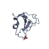
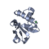
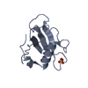
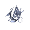
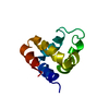
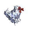
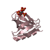
| タイトル | Crystal structure of the C-terminal PH domain of pleckstrin in complex with D-myo-Ins(1,2,3,4,5)P5 |
|---|
 要素 要素 | Pleckstrin |
|---|
 キーワード キーワード | LIPID BINDING PROTEIN / PH domain / protein-inositol phosphate complex |
|---|
| 機能・相同性 |  機能・相同性情報 機能・相同性情報
: / protein secretion by platelet / regulation of cell diameter / negative regulation of inositol phosphate biosynthetic process / positive regulation of actin filament depolymerization / platelet degranulation / phospholipase C-inhibiting G protein-coupled receptor signaling pathway / positive regulation of actin filament bundle assembly / positive regulation of integrin activation / cell projection organization ...: / protein secretion by platelet / regulation of cell diameter / negative regulation of inositol phosphate biosynthetic process / positive regulation of actin filament depolymerization / platelet degranulation / phospholipase C-inhibiting G protein-coupled receptor signaling pathway / positive regulation of actin filament bundle assembly / positive regulation of integrin activation / cell projection organization / ruffle organization / negative regulation of calcium-mediated signaling / phosphatidylinositol-3,4-bisphosphate binding / positive regulation of platelet activation / vesicle docking involved in exocytosis / cortical actin cytoskeleton organization / negative regulation of G protein-coupled receptor signaling pathway / thrombin-activated receptor signaling pathway / negative regulation of thrombin-activated receptor signaling pathway / hematopoietic progenitor cell differentiation / protein kinase C binding / integrin-mediated signaling pathway / platelet aggregation / ruffle membrane / Platelet degranulation / actin cytoskeleton organization / phospholipase C-activating G protein-coupled receptor signaling pathway / intracellular signal transduction / protein homodimerization activity / extracellular region / membrane / plasma membrane / cytoplasm / cytosol類似検索 - 分子機能 Pleckstrin, DEP domain / : / Domain found in Dishevelled, Egl-10, and Pleckstrin (DEP) / DEP domain profile. / Domain found in Dishevelled, Egl-10, and Pleckstrin / DEP domain / Pleckstrin-homology domain (PH domain)/Phosphotyrosine-binding domain (PTB) / PH-domain like / PH domain / PH domain profile. ...Pleckstrin, DEP domain / : / Domain found in Dishevelled, Egl-10, and Pleckstrin (DEP) / DEP domain profile. / Domain found in Dishevelled, Egl-10, and Pleckstrin / DEP domain / Pleckstrin-homology domain (PH domain)/Phosphotyrosine-binding domain (PTB) / PH-domain like / PH domain / PH domain profile. / Pleckstrin homology domain. / Pleckstrin homology domain / PH-like domain superfamily / Roll / Winged helix DNA-binding domain superfamily / Winged helix-like DNA-binding domain superfamily / Mainly Beta類似検索 - ドメイン・相同性 |
|---|
| 生物種 |  Homo sapiens (ヒト) Homo sapiens (ヒト) |
|---|
| 手法 |  X線回折 / シンクロトロン / 分子置換 / 解像度: 1.75 Å X線回折 / シンクロトロン / 分子置換 / 解像度: 1.75 Å |
|---|
 データ登録者 データ登録者 | Jackson, S.G. / Haslam, R.J. / Junop, M.S. |
|---|
 引用 引用 | ジャーナル: Bmc Struct.Biol. / 年: 2007
タイトル: Structural analysis of the carboxy terminal PH domain of pleckstrin bound to D-myo-inositol 1,2,3,5,6-pentakisphosphate.
著者: Jackson, S.G. / Zhang, Y. / Haslam, R.J. / Junop, M.S. |
|---|
| 履歴 | | 登録 | 2006年8月24日 | 登録サイト: RCSB / 処理サイト: RCSB |
|---|
| 改定 1.0 | 2007年8月7日 | Provider: repository / タイプ: Initial release |
|---|
| 改定 1.1 | 2011年7月13日 | Group: Version format compliance |
|---|
| 改定 1.2 | 2024年2月21日 | Group: Data collection / Database references / Derived calculations
カテゴリ: chem_comp_atom / chem_comp_bond ...chem_comp_atom / chem_comp_bond / database_2 / struct_ref_seq_dif / struct_site
Item: _database_2.pdbx_DOI / _database_2.pdbx_database_accession ..._database_2.pdbx_DOI / _database_2.pdbx_database_accession / _struct_ref_seq_dif.details / _struct_site.pdbx_auth_asym_id / _struct_site.pdbx_auth_comp_id / _struct_site.pdbx_auth_seq_id |
|---|
|
|---|
 ムービー
ムービー コントローラー


 データを開く
 基本情報
基本情報 構造の表示
構造の表示 ダウンロードとリンク
 その他のダウンロード


 PDBj
PDBj

 集合体
集合体


 分子量: 580.055 Da / 分子数: 3 / 由来タイプ: 合成 / 式: C6H17O21P5
 分子量: 18.015 Da / 分子数: 313 / 由来タイプ: 天然 / 式: H2O
 試料調製
試料調製 / ビームライン: X26C / 波長: 0.9797 Å
 解析
解析