 Movie
Movie Controller

[English] 日本語
 Yorodumi
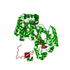
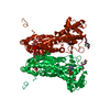
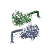
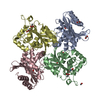
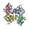
- PDB-2i1v: Crystal structure of PFKFB3 in complex with ADP and Fructose-2,6-... -
+ Open data

- Basic information
| Entry | Database: PDB / ID: 2i1v | |||||||||
|---|---|---|---|---|---|---|---|---|---|---|
| Title | Crystal structure of PFKFB3 in complex with ADP and Fructose-2,6-bisphosphate | |||||||||
 Components Components | 6-phosphofructo-2-kinase/fructose-2,6-biphosphatase 3 | |||||||||
 Keywords Keywords | TRANSFERASE / HYDROLASE / ternary product complex / ADP and F-2 / 6-P2 in 2-Kase / E-P and F-6-P in 2-Pase | |||||||||
| Function / homology |  Function and homology information Function and homology information6-phosphofructo-2-kinase/fructose-2,6-biphosphatase complex / 6-phosphofructo-2-kinase / 6-phosphofructo-2-kinase activity / fructose 2,6-bisphosphate metabolic process / fructose-2,6-bisphosphate 2-phosphatase / fructose-2,6-bisphosphate 2-phosphatase activity / Regulation of glycolysis by fructose 2,6-bisphosphate metabolism / glial cell differentiation / fructose metabolic process / motor behavior ...6-phosphofructo-2-kinase/fructose-2,6-biphosphatase complex / 6-phosphofructo-2-kinase / 6-phosphofructo-2-kinase activity / fructose 2,6-bisphosphate metabolic process / fructose-2,6-bisphosphate 2-phosphatase / fructose-2,6-bisphosphate 2-phosphatase activity / Regulation of glycolysis by fructose 2,6-bisphosphate metabolism / glial cell differentiation / fructose metabolic process / motor behavior / glycolytic process / gluconeogenesis / apoptotic process / nucleoplasm / ATP binding / cytosol Similarity search - Function | |||||||||
| Biological species |  Homo sapiens (human) Homo sapiens (human) | |||||||||
| Method |  X-RAY DIFFRACTION / SYNCHROTRON / MOLECULAR REPLACEMENT / Resolution: 2.5 Å X-RAY DIFFRACTION / SYNCHROTRON / MOLECULAR REPLACEMENT / Resolution: 2.5 Å | |||||||||
 Authors Authors | Kim, S.G. / El-Maghrabi, M.R. / Lee, Y.H. | |||||||||
 Citation Citation | Journal: J.Mol.Biol. / Year: 2007 Title: A Direct Substrate-Substrate Interaction Found in the Kinase Domain of the Bifunctional Enzyme, 6-Phosphofructo-2-kinase/Fructose-2,6-bisphosphatase Authors: Kim, S.G. / Cavalier, M. / El-Maghrabi, M.R. / Lee, Y.H. | |||||||||
| History |
|
- Structure visualization
| Structure viewer | Molecule: MolmilJmol/JSmol |
|---|
- Downloads & links
-Download
| PDBx/mmCIF format | 2i1v.cif.gz | 113.8 KB | Display | PDBx/mmCIF format |
|---|---|---|---|---|
| PDB format | pdb2i1v.ent.gz | 85.2 KB | Display | PDB format |
| PDBx/mmJSON format | 2i1v.json.gz | Tree view | PDBx/mmJSON format | |
| Others |  Other downloads Other downloads |
-Validation report
| Arichive directory | https://data.pdbj.org/pub/pdb/validation_reports/i1/2i1vftp://data.pdbj.org/pub/pdb/validation_reports/i1/2i1v | HTTPS FTP |
|---|
-Related structure data
| Related structure data |  2dwoC  2dwpC  2axnS S: Starting model for refinement C: citing same article ( |
|---|---|
| Similar structure data |

-Links
 PDBj
PDBj


- Assembly
| Deposited unit | 
| |||||||||
|---|---|---|---|---|---|---|---|---|---|---|
| 1 | 
| |||||||||
| Unit cell |
| |||||||||
| Components on special symmetry positions |
|
-Components
-Protein , 1 types, 1 molecules B
| #1: Protein | Mass: 59694.137 Da / Num. of mol.: 1 Source method: isolated from a genetically manipulated source Source: (gene. exp.) Homo sapiens (human) / Production host:  References: UniProt: Q16875, 6-phosphofructo-2-kinase, fructose-2,6-bisphosphate 2-phosphatase |
|---|
-Sugars , 2 types, 2 molecules 

| #2: Sugar | ChemComp-FDP /  Type: D-saccharide, beta linking / Mass: 340.116 Da / Num. of mol.: 1 Type: D-saccharide, beta linking / Mass: 340.116 Da / Num. of mol.: 1Source method: isolated from a genetically manipulated source Formula: C6H14O12P2 |
|---|---|
| #3: Sugar | ChemComp-F6P /  Type: D-saccharide, beta linking / Mass: 260.136 Da / Num. of mol.: 1 Type: D-saccharide, beta linking / Mass: 260.136 Da / Num. of mol.: 1Source method: isolated from a genetically manipulated source Formula: C6H13O9P |
-Non-polymers , 3 types, 224 molecules 

| #4: Chemical | ChemComp-ADP /  Mass: 427.201 Da / Num. of mol.: 1 / Source method: obtained synthetically / Formula: C10H15N5O10P2 / Comment: ADP, energy-carrying molecule*YM Mass: 427.201 Da / Num. of mol.: 1 / Source method: obtained synthetically / Formula: C10H15N5O10P2 / Comment: ADP, energy-carrying molecule*YM |
|---|---|
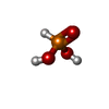
| #5: Chemical | ChemComp-PHS /  Mass: 81.996 Da / Num. of mol.: 1 / Source method: obtained synthetically / Formula: H3O3P Mass: 81.996 Da / Num. of mol.: 1 / Source method: obtained synthetically / Formula: H3O3P |
| #6: Water | ChemComp-HOH / Mass: 18.015 Da / Num. of mol.: 222 / Source method: isolated from a natural source / Formula: H2O |
-Experimental details
-Experiment
| Experiment | Method: X-RAY DIFFRACTION / Number of used crystals: 1 |
|---|
- Sample preparation
| Crystal | Density Matthews: 3.27 Å3/Da / Density % sol: 62.39 % |
|---|---|
| Crystal grow | Temperature: 298 K / Method: vapor diffusion, sitting drop / pH: 7.5 Details: PEG 4000, ethylene glycol, Tris, phosphate, pH 7.5, VAPOR DIFFUSION, SITTING DROP, temperature 298K |
-Data collection
| Diffraction | Mean temperature: 100 K |
|---|---|
| Diffraction source | Source: SYNCHROTRON / Site: CAMD  / Beamline: GCPCC / Wavelength: 1.3808 Å / Beamline: GCPCC / Wavelength: 1.3808 Å |
| Detector | Type: MAR CCD 165 mm / Detector: CCD / Date: Apr 13, 2005 |
| Radiation | Monochromator: SAGITALLY FOCUSED Si(111) / Protocol: SINGLE WAVELENGTH / Monochromatic (M) / Laue (L): M / Scattering type: x-ray |
| Radiation wavelength | Wavelength: 1.3808 Å / Relative weight: 1 |
| Reflection | Resolution: 2.5→30 Å / Num. obs: 28580 / % possible obs: 99.6 % / Observed criterion σ(F): 1 / Observed criterion σ(I): 2 / Redundancy: 13.9 % / Biso Wilson estimate: 47 Å2 / Rmerge(I) obs: 0.11 / Net I/σ(I): 15.3 |
| Reflection shell | Resolution: 2.5→2.59 Å / Redundancy: 1.9 % / Rmerge(I) obs: 0.535 / Mean I/σ(I) obs: 1.9 / Num. unique all: 2399 / % possible all: 98.2 |
- Processing
| Software |
| |||||||||||||||||||||||||
|---|---|---|---|---|---|---|---|---|---|---|---|---|---|---|---|---|---|---|---|---|---|---|---|---|---|---|
| Refinement | Method to determine structure: MOLECULAR REPLACEMENT Starting model: 2AXN Resolution: 2.5→30 Å / Isotropic thermal model: Isotropic / Cross valid method: THROUGHOUT / σ(F): 0 / Stereochemistry target values: Engh & Huber
| |||||||||||||||||||||||||
| Displacement parameters | Biso mean: 43.7 Å2 | |||||||||||||||||||||||||
| Refinement step | Cycle: LAST / Resolution: 2.5→30 Å
| |||||||||||||||||||||||||
| Refine LS restraints |
| |||||||||||||||||||||||||
| LS refinement shell | Resolution: 2.5→2.59 Å |
