 Movie
Movie Controller

[English] 日本語
 Yorodumi
- PDB-2hx7: Crystal structure of Cu(II) Azurin with the metal-binding loop se... -
+ Open data

- Basic information
| Entry | Database: PDB / ID: 2hx7 | ||||||
|---|---|---|---|---|---|---|---|
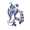
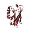
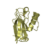
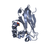
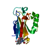
| Title | Crystal structure of Cu(II) Azurin with the metal-binding loop sequence "CTFPGHSALM" replaced with "CSPHQGAGM" | ||||||
 Components Components | Azurin | ||||||
 Keywords Keywords | ELECTRON TRANSPORT / BLUE COPPER-BINDING PROTEIN / GREEK-KEY BETA-BARREL / LOOP MUTAGENESIS | ||||||
| Function / homology |  Function and homology information Function and homology informationtransition metal ion binding / periplasmic space / electron transfer activity / copper ion binding / zinc ion binding / identical protein binding / plasma membrane Similarity search - Function | ||||||
| Biological species |   Pseudomonas aeruginosa (bacteria) Pseudomonas aeruginosa (bacteria) | ||||||
| Method |  X-RAY DIFFRACTION / MOLECULAR REPLACEMENT / Resolution: 1.55 Å X-RAY DIFFRACTION / MOLECULAR REPLACEMENT / Resolution: 1.55 Å | ||||||
 Authors Authors | Banfield, M.J. | ||||||
 Citation Citation | Journal: J.Am.Chem.Soc. / Year: 2007 Title: Engineering Copper Sites in Proteins: Loops Confer Native Structures and Properties to Chimeric Cupredoxins. Authors: Li, C. / Banfield, M.J. / Dennison, C. #1: Journal: Proc.Natl.Acad.Sci.USA / Year: 2006Title: Basic requirements for a metal-binding site in a protein: The influence of loop shorteneing on the cupredoxin azurin Authors: Chan, L. / Yanagisawa, S. / Martins, B.M. / Messerschmidt, A. / Banfield, M.J. / Dennison, C. | ||||||
| History |
|
- Structure visualization
| Structure viewer | Molecule: MolmilJmol/JSmol |
|---|
- Downloads & links
-Download
| PDBx/mmCIF format | 2hx7.cif.gz | 126 KB | Display | PDBx/mmCIF format |
|---|---|---|---|---|
| PDB format | pdb2hx7.ent.gz | 97.5 KB | Display | PDB format |
| PDBx/mmJSON format | 2hx7.json.gz | Tree view | PDBx/mmJSON format | |
| Others |  Other downloads Other downloads |
-Validation report
| Summary document | 2hx7_validation.pdf.gz | 432.8 KB | Display | wwPDB validaton report |
|---|---|---|---|---|
| Full document | 2hx7_full_validation.pdf.gz | 433.5 KB | Display | |
| Data in XML | 2hx7_validation.xml.gz | 16.5 KB | Display | |
| Data in CIF | 2hx7_validation.cif.gz | 25.6 KB | Display | |
| Arichive directory | https://data.pdbj.org/pub/pdb/validation_reports/hx/2hx7ftp://data.pdbj.org/pub/pdb/validation_reports/hx/2hx7 | HTTPS FTP |
-Related structure data
| Related structure data |  2hx8C  2hx9C  2hxaC  4azuS S: Starting model for refinement C: citing same article ( |
|---|---|
| Similar structure data |


-Links
 PDBj
PDBj- Assembly
| Deposited unit | 
| ||||||||
|---|---|---|---|---|---|---|---|---|---|
| 1 | 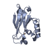
| ||||||||
| 2 | 
| ||||||||
| Unit cell |
|
-Components
| #1: Protein | Mass: 13785.544 Da / Num. of mol.: 2 Mutation: Metal binding loop "CTFPGHSALM" mutated to "CSPHQGAGM" Source method: isolated from a genetically manipulated source Source: (gene. exp.) Pseudomonas aeruginosa (bacteria) / Gene: azu / Plasmid: TRK99A / Production host: #2: Chemical |   Mass: 63.546 Da / Num. of mol.: 2 / Source method: obtained synthetically / Formula: Cu Mass: 63.546 Da / Num. of mol.: 2 / Source method: obtained synthetically / Formula: Cu#3: Water | ChemComp-HOH / |  Mass: 18.015 Da / Num. of mol.: 470 / Source method: isolated from a natural source / Formula: H2O Mass: 18.015 Da / Num. of mol.: 470 / Source method: isolated from a natural source / Formula: H2OHas protein modification | Y | |
|---|
-Experimental details
-Experiment
| Experiment | Method: X-RAY DIFFRACTION / Number of used crystals: 1 |
|---|
- Sample preparation
| Crystal | Density Matthews: 2.26 Å3/Da / Density % sol: 45.55 % |
|---|---|
| Crystal grow | Temperature: 293 K / Method: vapor diffusion, hanging drop / pH: 5 Details: 29-31% PEG 4000, 100mM magnesium chloride, 100mM Sodium acetate, pH 5, VAPOR DIFFUSION, HANGING DROP, temperature 293K |
-Data collection
| Diffraction | Mean temperature: 93 K |
|---|---|
| Diffraction source | Source: ROTATING ANODE / Type: RIGAKU / Wavelength: 1.542 Å |
| Detector | Type: RIGAKU RAXIS IV / Detector: IMAGE PLATE / Date: Mar 8, 2006 / Details: Osmic "blue" |
| Radiation | Monochromator: Ni FILTER / Protocol: SINGLE WAVELENGTH / Monochromatic (M) / Laue (L): M / Scattering type: x-ray |
| Radiation wavelength | Wavelength: 1.542 Å / Relative weight: 1 |
| Reflection | Resolution: 1.54→54.313 Å / Num. obs: 36537 / % possible obs: 96.6 % / Observed criterion σ(F): 0 / Observed criterion σ(I): 0 / Redundancy: 4.3 % / Biso Wilson estimate: 17.4 Å2 / Rmerge(I) obs: 0.051 / Rsym value: 0.051 / Net I/σ(I): 9.6 |
| Reflection shell | Resolution: 1.54→1.62 Å / Redundancy: 4.1 % / Rmerge(I) obs: 0.341 / Mean I/σ(I) obs: 2 / Num. measured all: 19172 / Num. unique all: 4673 / Rsym value: 0.341 / % possible all: 86 |
-Phasing
| Phasing MR |
|
|---|
- Processing
| Software |
| |||||||||||||||||||||||||||||||||||||||||||||||||||||||||||||||||||||||||||||||||||||||||||||||||||||||||||||||||||||||||||||||||||||||||||||||||
|---|---|---|---|---|---|---|---|---|---|---|---|---|---|---|---|---|---|---|---|---|---|---|---|---|---|---|---|---|---|---|---|---|---|---|---|---|---|---|---|---|---|---|---|---|---|---|---|---|---|---|---|---|---|---|---|---|---|---|---|---|---|---|---|---|---|---|---|---|---|---|---|---|---|---|---|---|---|---|---|---|---|---|---|---|---|---|---|---|---|---|---|---|---|---|---|---|---|---|---|---|---|---|---|---|---|---|---|---|---|---|---|---|---|---|---|---|---|---|---|---|---|---|---|---|---|---|---|---|---|---|---|---|---|---|---|---|---|---|---|---|---|---|---|---|---|---|
| Refinement | Method to determine structure: MOLECULAR REPLACEMENT Starting model: PDB entry 4AZU, with residues 112-121 removed Resolution: 1.55→30 Å / Cor.coef. Fo:Fc: 0.97 / Cor.coef. Fo:Fc free: 0.953 / SU B: 2.812 / SU ML: 0.047 / Cross valid method: THROUGHOUT / σ(F): 0 / ESU R: 0.098 / ESU R Free: 0.08 / Stereochemistry target values: MAXIMUM LIKELIHOOD Details: HYDROGENS HAVE BEEN ADDED IN THE RIDING POSITIONS, UT NOT OUTPUT TO THE FILE
| |||||||||||||||||||||||||||||||||||||||||||||||||||||||||||||||||||||||||||||||||||||||||||||||||||||||||||||||||||||||||||||||||||||||||||||||||
| Solvent computation | Ion probe radii: 0.8 Å / Shrinkage radii: 0.8 Å / VDW probe radii: 1.4 Å / Solvent model: MASK | |||||||||||||||||||||||||||||||||||||||||||||||||||||||||||||||||||||||||||||||||||||||||||||||||||||||||||||||||||||||||||||||||||||||||||||||||
| Displacement parameters | Biso mean: 9.984 Å2
| |||||||||||||||||||||||||||||||||||||||||||||||||||||||||||||||||||||||||||||||||||||||||||||||||||||||||||||||||||||||||||||||||||||||||||||||||
| Refinement step | Cycle: LAST / Resolution: 1.55→30 Å
| |||||||||||||||||||||||||||||||||||||||||||||||||||||||||||||||||||||||||||||||||||||||||||||||||||||||||||||||||||||||||||||||||||||||||||||||||
| Refine LS restraints |
| |||||||||||||||||||||||||||||||||||||||||||||||||||||||||||||||||||||||||||||||||||||||||||||||||||||||||||||||||||||||||||||||||||||||||||||||||
| LS refinement shell | Resolution: 1.55→1.59 Å / Total num. of bins used: 20
|
