 Movie
Movie Controller

[English] 日本語
 Yorodumi
- PDB-2hug: 3D Solution Structure of the Chromo-2 Domain of cpSRP43 complexed... -
+ Open data

- Basic information
| Entry | Database: PDB / ID: 2hug | ||||||
|---|---|---|---|---|---|---|---|
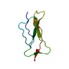
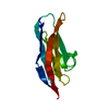
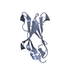
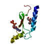
| Title | 3D Solution Structure of the Chromo-2 Domain of cpSRP43 complexed with cpSRP54 peptide | ||||||
 Components Components |
| ||||||
 Keywords Keywords | PLANT PROTEIN / Chromo-2 domain / cpSRP43 / LHCP / thylakoid / protein translocation / cpSRP54 | ||||||
| Function / homology |  Function and homology information Function and homology informationprotein import into chloroplast thylakoid membrane / protein heterotrimerization / signal recognition particle, endoplasmic reticulum targeting / response to high light intensity / signal-recognition-particle GTPase / 7S RNA binding / SRP-dependent cotranslational protein targeting to membrane / chloroplast envelope / chloroplast stroma / chloroplast thylakoid membrane ...protein import into chloroplast thylakoid membrane / protein heterotrimerization / signal recognition particle, endoplasmic reticulum targeting / response to high light intensity / signal-recognition-particle GTPase / 7S RNA binding / SRP-dependent cotranslational protein targeting to membrane / chloroplast envelope / chloroplast stroma / chloroplast thylakoid membrane / chloroplast / disordered domain specific binding / protein-macromolecule adaptor activity / protein domain specific binding / GTPase activity / GTP binding / protein-containing complex / metal ion binding / identical protein binding / plasma membrane / cytosol Similarity search - Function | ||||||
| Biological species |  | ||||||
| Method | SOLUTION NMR / distance geometry, simulated annealing, molecular dynamics, torsion angle dynamics | ||||||
 Authors Authors | Kathir, K.M. / Vaithiyalingam, S. / Henry, R. / Thallapuranam, S.K.K. | ||||||
 Citation Citation | Journal: J.Mol.Biol. / Year: 2008 Title: Assembly of chloroplast signal recognition particle involves structural rearrangement in cpSRP43. Authors: Kathir, K.M. / Rajalingam, D. / Sivaraja, V. / Kight, A. / Goforth, R.L. / Yu, C. / Henry, R. / Kumar, T.K. | ||||||
| History |
|
- Structure visualization
| Structure viewer | Molecule: MolmilJmol/JSmol |
|---|
- Downloads & links
-Download
| PDBx/mmCIF format | 2hug.cif.gz | 483.1 KB | Display | PDBx/mmCIF format |
|---|---|---|---|---|
| PDB format | pdb2hug.ent.gz | 407.1 KB | Display | PDB format |
| PDBx/mmJSON format | 2hug.json.gz | Tree view | PDBx/mmJSON format | |
| Others |  Other downloads Other downloads |
-Validation report
| Arichive directory | https://data.pdbj.org/pub/pdb/validation_reports/hu/2hugftp://data.pdbj.org/pub/pdb/validation_reports/hu/2hug | HTTPS FTP |
|---|
-Related structure data
| Similar structure data | |
|---|---|
| Other databases |


-Links
 PDBj
PDBj- Assembly
| Deposited unit | 
| |||||||||
|---|---|---|---|---|---|---|---|---|---|---|
| 1 |
| |||||||||
| NMR ensembles |
|
-Components
| #1: Protein | Mass: 6548.151 Da / Num. of mol.: 1 / Fragment: Chromo-2 domain (residues 265-319) Source method: isolated from a genetically manipulated source Source: (gene. exp.)  |
|---|---|
| #2: Protein/peptide | Mass: 1515.740 Da / Num. of mol.: 1 / Fragment: M-domain (residues 530-543) Source method: isolated from a genetically manipulated source Source: (gene. exp.) |
-Experimental details
-Experiment
| Experiment | Method: SOLUTION NMR |
|---|
- Sample preparation
| Details | Contents: 1.5mM Chromo domain-2;Uniform labeling with 13C, 15N at known labeling levels: U-95% 13C;U-98% 15N; PBS buffer. Solvent system: 90% H2O/10% D2O |
|---|---|
| Sample conditions | Ionic strength: 150 mM NaCl / pH: 6.5 / Pressure: ambient / Temperature: 298 K |
-NMR measurement
| Radiation | Protocol: SINGLE WAVELENGTH / Monochromatic (M) / Laue (L): M |
|---|---|
| Radiation wavelength | Relative weight: 1 |
| NMR spectrometer | Type: Bruker AVANCE / Manufacturer: Bruker / Model: AVANCE / Field strength: 700 MHz |
- Processing
| NMR software |
| ||||||||||||
|---|---|---|---|---|---|---|---|---|---|---|---|---|---|
| Refinement | Method: distance geometry, simulated annealing, molecular dynamics, torsion angle dynamics Software ordinal: 1 | ||||||||||||
| NMR representative | Selection criteria: lowest energy | ||||||||||||
| NMR ensemble | Conformers calculated total number: 200 / Conformers submitted total number: 20 |
