 Movie
Movie Controller


+ Open data

- Basic information
| Entry | Database: PDB / ID: 2hdd | ||||||
|---|---|---|---|---|---|---|---|
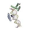
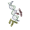
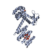
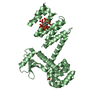
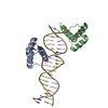
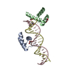
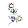
| Title | ENGRAILED HOMEODOMAIN Q50K VARIANT DNA COMPLEX | ||||||
 Components Components |
| ||||||
 Keywords Keywords | TRANSCRIPTION/DNA / DNA BINDING / COMPLEX (DNA BINDING PROTEIN-DNA) / TRANSCRIPTION-DNA COMPLEX | ||||||
| Function / homology |  Function and homology information Function and homology informationposterior compartment specification / analia development / anterior head segmentation / anterior/posterior lineage restriction, imaginal disc / genital disc development / genital disc anterior/posterior pattern formation / posterior head segmentation / trunk segmentation / spiracle morphogenesis, open tracheal system / wing disc anterior/posterior pattern formation ...posterior compartment specification / analia development / anterior head segmentation / anterior/posterior lineage restriction, imaginal disc / genital disc development / genital disc anterior/posterior pattern formation / posterior head segmentation / trunk segmentation / spiracle morphogenesis, open tracheal system / wing disc anterior/posterior pattern formation / wing disc morphogenesis / imaginal disc-derived wing vein specification / neuroblast fate determination / segment polarity determination / ventral midline development / compartment pattern specification / gonad development / axon guidance / RNA polymerase II transcription regulatory region sequence-specific DNA binding / DNA-binding transcription repressor activity, RNA polymerase II-specific / neuron differentiation / regulation of gene expression / sequence-specific DNA binding / negative regulation of neuron apoptotic process / DNA-binding transcription factor activity, RNA polymerase II-specific / RNA polymerase II cis-regulatory region sequence-specific DNA binding / negative regulation of gene expression / regulation of transcription by RNA polymerase II / negative regulation of transcription by RNA polymerase II / positive regulation of transcription by RNA polymerase II / nucleus Similarity search - Function | ||||||
| Biological species |  | ||||||
| Method |  X-RAY DIFFRACTION / MIRAS / Resolution: 1.9 Å X-RAY DIFFRACTION / MIRAS / Resolution: 1.9 Å | ||||||
 Authors Authors | Tucker-Kellogg, L. / Rould, M.A. / Chambers, K.A. / Ades, S.E. / Sauer, R.T. / Pabo, C.O. | ||||||
 Citation Citation | Journal: Structure / Year: 1997 Title: Engrailed (Gln50-->Lys) homeodomain-DNA complex at 1.9 A resolution: structural basis for enhanced affinity and altered specificity. Authors: Tucker-Kellogg, L. / Rould, M.A. / Chambers, K.A. / Ades, S.E. / Sauer, R.T. / Pabo, C.O. | ||||||
| History |
|
- Structure visualization
| Structure viewer | Molecule: MolmilJmol/JSmol |
|---|
- Downloads & links
-Download
| PDBx/mmCIF format | 2hdd.cif.gz | 63.2 KB | Display | PDBx/mmCIF format |
|---|---|---|---|---|
| PDB format | pdb2hdd.ent.gz | 44.2 KB | Display | PDB format |
| PDBx/mmJSON format | 2hdd.json.gz | Tree view | PDBx/mmJSON format | |
| Others |  Other downloads Other downloads |
-Validation report
| Arichive directory | https://data.pdbj.org/pub/pdb/validation_reports/hd/2hddftp://data.pdbj.org/pub/pdb/validation_reports/hd/2hdd | HTTPS FTP |
|---|
-Related structure data
| Similar structure data |
|---|

-Links
 PDBj
PDBj


- Assembly
| Deposited unit | 
| ||||||||
|---|---|---|---|---|---|---|---|---|---|
| 1 |
| ||||||||
| Unit cell |
| ||||||||
| Noncrystallographic symmetry (NCS) | NCS oper: (Code: given Matrix: (-0.703806, -0.704302, 0.092822), Vector: |
-Components
| #1: DNA chain | Mass: 6389.133 Da / Num. of mol.: 1 / Source method: obtained synthetically | ||
|---|---|---|---|
| #2: DNA chain | Mass: 6496.224 Da / Num. of mol.: 1 / Source method: obtained synthetically | ||
| #3: Protein | Mass: 7426.592 Da / Num. of mol.: 2 / Mutation: Q50K Source method: isolated from a genetically manipulated source Source: (gene. exp.)  #4: Water | ChemComp-HOH / |  Mass: 18.015 Da / Num. of mol.: 183 / Source method: isolated from a natural source / Formula: H2O Mass: 18.015 Da / Num. of mol.: 183 / Source method: isolated from a natural source / Formula: H2O |
-Experimental details
-Experiment
| Experiment | Method: X-RAY DIFFRACTION / Number of used crystals: 1 |
|---|
- Sample preparation
| Crystal | Density Matthews: 3.23 Å3/Da / Density % sol: 48 % | |||||||||||||||||||||||||||||||||||
|---|---|---|---|---|---|---|---|---|---|---|---|---|---|---|---|---|---|---|---|---|---|---|---|---|---|---|---|---|---|---|---|---|---|---|---|---|
| Crystal grow | Method: vapor diffusion, hanging drop / pH: 7 Details: COMPLEX WAS CRYSTALLIZED BY HANGING DROP VAPOR DIFFUSION OVER .75M AMOAC AND 1% PEG400; DROP STARTS AT 1M AMOAC., pH 7.00, vapor diffusion - hanging drop | |||||||||||||||||||||||||||||||||||
| Components of the solutions |
| |||||||||||||||||||||||||||||||||||
| Crystal | *PLUS Density % sol: 48 % | |||||||||||||||||||||||||||||||||||
| Crystal grow | *PLUS | |||||||||||||||||||||||||||||||||||
| Components of the solutions | *PLUS
|
-Data collection
| Diffraction | Mean temperature: 123 K |
|---|---|
| Diffraction source | Source: ROTATING ANODE / Type: RIGAKU RU200 |
| Detector | Type: RIGAKU RAXIS IIC / Detector: IMAGE PLATE / Date: Jun 1, 1995 / Details: MIRRORS |
| Radiation | Monochromator: NI MIRROR + NI FILTER / Monochromatic (M) / Laue (L): M / Scattering type: x-ray |
| Radiation wavelength | Relative weight: 1 |
| Reflection | Resolution: 1.9→20 Å / Num. obs: 27136 / % possible obs: 94 % / Redundancy: 3.4 % / Rmerge(I) obs: 0.035 / Rsym value: 0.044 |
| Reflection shell | Resolution: 1.9→1.97 Å / Rmerge(I) obs: 0.38 / % possible all: 84 |
| Reflection | *PLUS Highest resolution: 1.9 Å / Lowest resolution: 20 Å / % possible obs: 94 % / Redundancy: 3.4 % |
| Reflection shell | *PLUS Highest resolution: 1.9 Å / Lowest resolution: 1.97 Å / % possible obs: 94 % / Mean I/σ(I) obs: 2.2 |
- Processing
| Software |
| ||||||||||||||||||||||||||||||||||||||||||||||||||||||||||||
|---|---|---|---|---|---|---|---|---|---|---|---|---|---|---|---|---|---|---|---|---|---|---|---|---|---|---|---|---|---|---|---|---|---|---|---|---|---|---|---|---|---|---|---|---|---|---|---|---|---|---|---|---|---|---|---|---|---|---|---|---|---|
| Refinement | Method to determine structure: MIRAS / Resolution: 1.9→6 Å / Data cutoff high absF: 1000000000 / Data cutoff low absF: 0 / Isotropic thermal model: RESTRAINED / Cross valid method: THROUGHOUT / σ(F): 2
| ||||||||||||||||||||||||||||||||||||||||||||||||||||||||||||
| Refinement step | Cycle: LAST / Resolution: 1.9→6 Å
| ||||||||||||||||||||||||||||||||||||||||||||||||||||||||||||
| Refine LS restraints |
| ||||||||||||||||||||||||||||||||||||||||||||||||||||||||||||
| LS refinement shell | Resolution: 1.9→1.97 Å / % reflection Rfree: 10 % / Total num. of bins used: 10 / % reflection obs: 84 % | ||||||||||||||||||||||||||||||||||||||||||||||||||||||||||||
| Xplor file |
| ||||||||||||||||||||||||||||||||||||||||||||||||||||||||||||
| Software | *PLUS Name: X-PLOR / Version: 3.1 / Classification: refinement | ||||||||||||||||||||||||||||||||||||||||||||||||||||||||||||
| Refinement | *PLUS Highest resolution: 1.9 Å / Lowest resolution: 6 Å / σ(F): 2 / % reflection Rfree: 10 % | ||||||||||||||||||||||||||||||||||||||||||||||||||||||||||||
| Solvent computation | *PLUS | ||||||||||||||||||||||||||||||||||||||||||||||||||||||||||||
| Displacement parameters | *PLUS |
