 Movie
Movie Controller


+ Open data

- Basic information
| Entry | Database: PDB / ID: 1va7 | ||||||
|---|---|---|---|---|---|---|---|
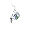
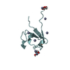
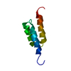
| Title | Yeast Myo3 SH3 domain, triclinic crystal form | ||||||
 Components Components | Myosin-3 isoform | ||||||
 Keywords Keywords | CONTRACTILE PROTEIN / Structural genomics / SH3 domain | ||||||
| Function / homology |  Function and homology information Function and homology informationbipolar cellular bud site selection / positive regulation of Arp2/3 complex-mediated actin nucleation / actin cortical patch localization / fungal-type cell wall organization / actin cortical patch / cell tip / myosin complex / reticulophagy / response to osmotic stress / myosin binding ...bipolar cellular bud site selection / positive regulation of Arp2/3 complex-mediated actin nucleation / actin cortical patch localization / fungal-type cell wall organization / actin cortical patch / cell tip / myosin complex / reticulophagy / response to osmotic stress / myosin binding / microfilament motor activity / exocytosis / microvillus / cell periphery / actin filament organization / endocytosis / actin filament binding / actin cytoskeleton / hydrolase activity / ATP binding / plasma membrane / cytoplasm Similarity search - Function | ||||||
| Biological species |  | ||||||
| Method |  X-RAY DIFFRACTION / SYNCHROTRON / MOLECULAR REPLACEMENT / Resolution: 2.9 Å X-RAY DIFFRACTION / SYNCHROTRON / MOLECULAR REPLACEMENT / Resolution: 2.9 Å | ||||||
 Authors Authors | Kursula, P. / Lehmann, F. / Song, Y.H. / Wilmanns, M. | ||||||
 Citation Citation | Journal: To be Published Title: High-throughput structural genomics of yeast SH3 domains Authors: Kursula, P. / Lehmann, F. / Song, Y.H. / Wilmanns, M. | ||||||
| History |
|
- Structure visualization
| Structure viewer | Molecule: MolmilJmol/JSmol |
|---|
- Downloads & links
-Download
| PDBx/mmCIF format | 1va7.cif.gz | 58.6 KB | Display | PDBx/mmCIF format |
|---|---|---|---|---|
| PDB format | pdb1va7.ent.gz | 43.4 KB | Display | PDB format |
| PDBx/mmJSON format | 1va7.json.gz | Tree view | PDBx/mmJSON format | |
| Others |  Other downloads Other downloads |
-Validation report
| Arichive directory | https://data.pdbj.org/pub/pdb/validation_reports/va/1va7ftp://data.pdbj.org/pub/pdb/validation_reports/va/1va7 | HTTPS FTP |
|---|
-Related structure data
| Related structure data | 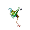 1ruwS S: Starting model for refinement |
|---|---|
| Similar structure data |


-Links
 PDBj
PDBj


- Assembly
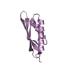
| Deposited unit | 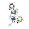
| ||||||||||||||||||||||||||||||
|---|---|---|---|---|---|---|---|---|---|---|---|---|---|---|---|---|---|---|---|---|---|---|---|---|---|---|---|---|---|---|---|
| 1 | 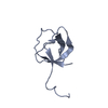
| ||||||||||||||||||||||||||||||
| 2 | 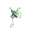
| ||||||||||||||||||||||||||||||
| 3 | 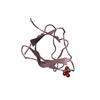
| ||||||||||||||||||||||||||||||
| 4 | 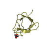
| ||||||||||||||||||||||||||||||
| Unit cell |
| ||||||||||||||||||||||||||||||
| Noncrystallographic symmetry (NCS) | NCS domain:
NCS domain segments: Component-ID: 1 / Ens-ID: 1 / Beg auth comp-ID: ASP / Beg label comp-ID: ASP / End auth comp-ID: TYR / End label comp-ID: TYR / Refine code: 1 / Auth seq-ID: 3 - 61 / Label seq-ID: 3 - 61
| ||||||||||||||||||||||||||||||
| Details | each chain is an independent biological unit |
-Components
| #1: Protein | Mass: 7645.547 Da / Num. of mol.: 4 / Fragment: SH3 domain Source method: isolated from a genetically manipulated source Source: (gene. exp.) Plasmid: pDEST17 / Production host:  #2: Chemical |   Mass: 92.094 Da / Num. of mol.: 2 / Source method: obtained synthetically / Formula: C3H8O3 Mass: 92.094 Da / Num. of mol.: 2 / Source method: obtained synthetically / Formula: C3H8O3 |
|---|
-Experimental details
-Experiment
| Experiment | Method: X-RAY DIFFRACTION / Number of used crystals: 1 |
|---|
- Sample preparation
| Crystal | Density Matthews: 2.67 Å3/Da / Density % sol: 53.49 % |
|---|---|
| Crystal grow | Temperature: 293 K / Method: vapor diffusion, sitting drop / pH: 7.5 Details: sodium citrate, glycerol, pH 7.5, VAPOR DIFFUSION, SITTING DROP, temperature 293K |
-Data collection
| Diffraction | Mean temperature: 100 K |
|---|---|
| Diffraction source | Source: SYNCHROTRON / Site: EMBL/DESY, HAMBURG  / Beamline: X11 / Wavelength: 0.8115 Å / Beamline: X11 / Wavelength: 0.8115 Å |
| Detector | Type: MARRESEARCH / Detector: CCD / Date: Feb 10, 2004 |
| Radiation | Protocol: SINGLE WAVELENGTH / Monochromatic (M) / Laue (L): M / Scattering type: x-ray |
| Radiation wavelength | Wavelength: 0.8115 Å / Relative weight: 1 |
| Reflection | Resolution: 2.9→20 Å / Num. all: 6221 / Num. obs: 6221 / % possible obs: 96.7 % / Observed criterion σ(F): -3 / Observed criterion σ(I): -3 / Redundancy: 2.1 % / Biso Wilson estimate: 33 Å2 / Rsym value: 0.106 / Net I/σ(I): 7.1 |
| Reflection shell | Resolution: 2.9→3 Å / Redundancy: 2.1 % / Mean I/σ(I) obs: 2.5 / Num. unique all: 609 / Rsym value: 0.406 / % possible all: 96.8 |
- Processing
| Software |
| |||||||||||||||||||||||||||||||||||||||||||||||||||||||||||||||||||||||||||||||||||||||||||||||||||||||||||||||||||||||||||||
|---|---|---|---|---|---|---|---|---|---|---|---|---|---|---|---|---|---|---|---|---|---|---|---|---|---|---|---|---|---|---|---|---|---|---|---|---|---|---|---|---|---|---|---|---|---|---|---|---|---|---|---|---|---|---|---|---|---|---|---|---|---|---|---|---|---|---|---|---|---|---|---|---|---|---|---|---|---|---|---|---|---|---|---|---|---|---|---|---|---|---|---|---|---|---|---|---|---|---|---|---|---|---|---|---|---|---|---|---|---|---|---|---|---|---|---|---|---|---|---|---|---|---|---|---|---|---|
| Refinement | Method to determine structure: MOLECULAR REPLACEMENT Starting model: 1RUW Resolution: 2.9→20 Å / Cor.coef. Fo:Fc: 0.897 / Cor.coef. Fo:Fc free: 0.846 / SU B: 24.02 / SU ML: 0.436 / TLS residual ADP flag: LIKELY RESIDUAL / Isotropic thermal model: TLS parameters used / Cross valid method: THROUGHOUT / σ(F): -3 / ESU R Free: 0.484 / Stereochemistry target values: Engh & Huber / Details: HYDROGENS HAVE BEEN ADDED IN THE RIDING POSITIONS
| |||||||||||||||||||||||||||||||||||||||||||||||||||||||||||||||||||||||||||||||||||||||||||||||||||||||||||||||||||||||||||||
| Solvent computation | Ion probe radii: 0.8 Å / Shrinkage radii: 0.8 Å / VDW probe radii: 1.4 Å / Solvent model: BABINET MODEL WITH MASK | |||||||||||||||||||||||||||||||||||||||||||||||||||||||||||||||||||||||||||||||||||||||||||||||||||||||||||||||||||||||||||||
| Displacement parameters | Biso mean: 6.195 Å2
| |||||||||||||||||||||||||||||||||||||||||||||||||||||||||||||||||||||||||||||||||||||||||||||||||||||||||||||||||||||||||||||
| Refinement step | Cycle: LAST / Resolution: 2.9→20 Å
| |||||||||||||||||||||||||||||||||||||||||||||||||||||||||||||||||||||||||||||||||||||||||||||||||||||||||||||||||||||||||||||
| Refine LS restraints |
| |||||||||||||||||||||||||||||||||||||||||||||||||||||||||||||||||||||||||||||||||||||||||||||||||||||||||||||||||||||||||||||
| Refine LS restraints NCS | Ens-ID: 1 / Number: 860 / Refine-ID: X-RAY DIFFRACTION
| |||||||||||||||||||||||||||||||||||||||||||||||||||||||||||||||||||||||||||||||||||||||||||||||||||||||||||||||||||||||||||||
| LS refinement shell | Resolution: 2.9→2.974 Å / Total num. of bins used: 20 /
| |||||||||||||||||||||||||||||||||||||||||||||||||||||||||||||||||||||||||||||||||||||||||||||||||||||||||||||||||||||||||||||
| Refinement TLS params. | Method: refined / Refine-ID: X-RAY DIFFRACTION
| |||||||||||||||||||||||||||||||||||||||||||||||||||||||||||||||||||||||||||||||||||||||||||||||||||||||||||||||||||||||||||||
| Refinement TLS group |
|
