SEQUENCE THE CONSTRUCT WAS EXPRESSED WITH A PURIFICATION TAG MGSDKIHHHHHHENLYFQG. THE TAG WAS ...SEQUENCE THE CONSTRUCT WAS EXPRESSED WITH A PURIFICATION TAG MGSDKIHHHHHHENLYFQG. THE TAG WAS REMOVED WITH TEV PROTEASE LEAVING ONLY A GLYCINE (0) FOLLOWED BY THE TARGET SEQUENCE.
Resolution: 1.8→27.951 Å / Num. obs: 21098 / % possible obs: 92.2 % / Biso Wilson estimate: 25.562 Å2 / Rmerge(I) obs: 0.069 / Net I/σ(I): 8.1
Reflection shell
Diffraction-ID: 1
Resolution (Å)
Highest resolution (Å)
Rmerge(I) obs
Mean I/σ(I) obs
Num. measured obs
Num. unique all
% possible all
1.8-1.86
0.477
1.6
4474
2718
72.7
1.86-1.94
0.33
2.3
6935
3654
84.3
1.94-2.03
0.266
2.7
6966
3551
85.8
2.03-2.13
0.201
3.8
6877
3541
93.4
2.13-2.27
0.132
5.3
7917
4077
96.8
2.27-2.44
0.106
6.5
7383
3818
97.7
2.44-2.69
0.093
7.3
7784
4038
97.2
2.69-3.07
0.066
9.3
7430
3810
96.6
3.07
0.044
14.4
7814
4015
98.1
-
Phasing
Phasing
Method: MAD
-
Processing
Software
Name
Version
Classification
NB
MolProbity
3beta29
modelbuilding
REFMAC
5.2.0005
refinement
XSCALE
datascaling
PDB_EXTRACT
2
dataextraction
XDS
datareduction
Refinement
Method to determine structure: MAD / Resolution: 1.8→27.951 Å / Cor.coef. Fo:Fc: 0.955 / Cor.coef. Fo:Fc free: 0.918 / SU B: 6.882 / SU ML: 0.11 / TLS residual ADP flag: LIKELY RESIDUAL / Cross valid method: THROUGHOUT / σ(F): 0 / ESU R: 0.142 / ESU R Free: 0.144 Stereochemistry target values: MAXIMUM LIKELIHOOD WITH PHASES Details: 1. HYDROGENS HAVE BEEN ADDED IN THE RIDING POSITIONS. 2. A MET-INHIBITION PROTOCOL WAS USED FOR SELENOMETHIONINE INCORPORATION DURING PROTEIN EXPRESSION. THE OCCUPANCY OF THE SE ATOMS IN THE ...Details: 1. HYDROGENS HAVE BEEN ADDED IN THE RIDING POSITIONS. 2. A MET-INHIBITION PROTOCOL WAS USED FOR SELENOMETHIONINE INCORPORATION DURING PROTEIN EXPRESSION. THE OCCUPANCY OF THE SE ATOMS IN THE MSE RESIDUES WAS REDUCED TO 0.75 FOR THE REDUCED SCATTERING POWER DUE TO PARTIAL S-MET INCORPORATION. 3. MG MODELED IN THE ACTIVE SITE BASED ON CRYSTALLIZATION CONDITIONS AND RELATED STRUCTURES. 4. MG MODELED NEAR GLU 63 BASED ON GEOMETRY AND CRYSTLLIZATION CONDITIONS. 5. CHLORINE MODELED BASED ON CRYSTALLIZATION CONDITIONS. 6. RESIDUES 40-42 WERE DISORDERED AND NOT MODELED. 7. ATOM RECORD CONTAINS RESIDUAL B FACTORS ONLY.
Rfactor
Num. reflection
% reflection
Selection details
Rfree
0.247
1084
5.1 %
RANDOM
Rwork
0.189
-
-
-
obs
0.192
21060
97.65 %
-
Solvent computation
Ion probe radii: 0.8 Å / Shrinkage radii: 0.8 Å / VDW probe radii: 1.2 Å / Solvent model: MASK
Displacement parameters
Biso mean: 19.764 Å2
Baniso -1
Baniso -2
Baniso -3
1-
2.23 Å2
0 Å2
0 Å2
2-
-
-0.52 Å2
0 Å2
3-
-
-
-1.7 Å2
Refinement step
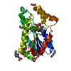
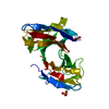
Cycle: LAST / Resolution: 1.8→27.951 Å
Protein
Nucleic acid
Ligand
Solvent
Total
Num. atoms
1744
0
3
235
1982
Refine LS restraints
Refine-ID
Type
Dev ideal
Dev ideal target
Number
X-RAY DIFFRACTION
r_bond_refined_d
0.015
0.022
1783
X-RAY DIFFRACTION
r_bond_other_d
0.001
0.02
1679
X-RAY DIFFRACTION
r_angle_refined_deg
1.366
1.976
2408
X-RAY DIFFRACTION
r_angle_other_deg
0.8
3
3872
X-RAY DIFFRACTION
r_dihedral_angle_1_deg
7.139
5
227
X-RAY DIFFRACTION
r_dihedral_angle_2_deg
34.064
23.294
85
X-RAY DIFFRACTION
r_dihedral_angle_3_deg
11.989
15
315
X-RAY DIFFRACTION
r_dihedral_angle_4_deg
18.147
15
18
X-RAY DIFFRACTION
r_chiral_restr
0.076
0.2
279
X-RAY DIFFRACTION
r_gen_planes_refined
0.006
0.02
2001
X-RAY DIFFRACTION
r_gen_planes_other
0.001
0.02
377
X-RAY DIFFRACTION
r_nbd_refined
0.211
0.2
379
X-RAY DIFFRACTION
r_nbd_other
0.181
0.2
1718
X-RAY DIFFRACTION
r_nbtor_refined
0.176
0.2
883
X-RAY DIFFRACTION
r_nbtor_other
0.084
0.2
1044
X-RAY DIFFRACTION
r_xyhbond_nbd_refined
0.163
0.2
154
X-RAY DIFFRACTION
r_metal_ion_refined
0.046
0.2
1
X-RAY DIFFRACTION
r_symmetry_vdw_refined
0.08
0.2
4
X-RAY DIFFRACTION
r_symmetry_vdw_other
0.244
0.2
34
X-RAY DIFFRACTION
r_symmetry_hbond_refined
0.171
0.2
17
X-RAY DIFFRACTION
r_mcbond_it
0.955
1.5
1230
X-RAY DIFFRACTION
r_mcbond_other
0.21
1.5
465
X-RAY DIFFRACTION
r_mcangle_it
1.268
2
1789
X-RAY DIFFRACTION
r_scbond_it
2.231
3
704
X-RAY DIFFRACTION
r_scangle_it
3.195
4.5
617
LS refinement shell
Resolution: 1.8→1.847 Å / Total num. of bins used: 20
Rfactor
Num. reflection
% reflection
Rfree
0.358
57
-
Rwork
0.297
1201
-
obs
-
1258
82.28 %
Refinement TLS params.
Method: refined / Origin x: 22.239 Å / Origin y: 44.9764 Å / Origin z: 15.7082 Å
11
12
13
21
22
23
31
32
33
T
-0.0707 Å2
0.0126 Å2
0.007 Å2
-
-0.0257 Å2
-0.0012 Å2
-
-
-0.0338 Å2
L
0.4396 °2
0.0601 °2
-0.021 °2
-
0.8482 °2
0.1049 °2
-
-
1.1383 °2
S
0.0059 Å °
-0.0257 Å °
0.0104 Å °
-0.0382 Å °
-0.0041 Å °
-0.0069 Å °
-0.0322 Å °
-0.0215 Å °
-0.0018 Å °
Refinement TLS group
Refine-ID: X-RAY DIFFRACTION / Refine TLS-ID: 1 / Selection: ALL / Auth asym-ID: A / Label asym-ID: A
ID
Auth seq-ID
Label seq-ID
1
2 - 39
3 - 40
2
43 - 229
44 - 230
+
About Yorodumi
-
News
-
Feb 9, 2022. New format data for meta-information of EMDB entries
New format data for meta-information of EMDB entries
Version 3 of the EMDB header file is now the official format.
The previous official version 1.9 will be removed from the archive.
In the structure databanks used in Yorodumi, some data are registered as the other names, "COVID-19 virus" and "2019-nCoV". Here are the details of the virus and the list of structure data.
Jan 31, 2019. EMDB accession codes are about to change! (news from PDBe EMDB page)
EMDB accession codes are about to change! (news from PDBe EMDB page)
The allocation of 4 digits for EMDB accession codes will soon come to an end. Whilst these codes will remain in use, new EMDB accession codes will include an additional digit and will expand incrementally as the available range of codes is exhausted. The current 4-digit format prefixed with “EMD-” (i.e. EMD-XXXX) will advance to a 5-digit format (i.e. EMD-XXXXX), and so on. It is currently estimated that the 4-digit codes will be depleted around Spring 2019, at which point the 5-digit format will come into force.
The EM Navigator/Yorodumi systems omit the EMD- prefix.
Related info.:Q: What is EMD? / ID/Accession-code notation in Yorodumi/EM Navigator
Yorodumi is a browser for structure data from EMDB, PDB, SASBDB, etc.
This page is also the successor to EM Navigator detail page, and also detail information page/front-end page for Omokage search.
The word "yorodu" (or yorozu) is an old Japanese word meaning "ten thousand". "mi" (miru) is to see.
Related info.:EMDB / PDB / SASBDB / Comparison of 3 databanks / Yorodumi Search / Aug 31, 2016. New EM Navigator & Yorodumi / Yorodumi Papers / Jmol/JSmol / Function and homology information / Changes in new EM Navigator and Yorodumi
 Movie
Movie Controller

 Yorodumi
 Open data
 Basic information
 Components
 Keywords
 Function and homology information
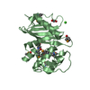
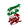
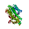
 Chlorobaculum tepidum (bacteria)
 X-RAY DIFFRACTION /
  Authors
 Citation
 Structure visualization
 Downloads & links
 Other downloads
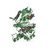

 PDBj
PDBj
 Assembly

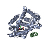
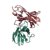
 Mass: 24.305 Da / Num. of mol.: 2 / Source method: obtained synthetically / Formula: Mg
 Mass: 35.453 Da / Num. of mol.: 1 / Source method: obtained synthetically / Formula: Cl
 Mass: 18.015 Da / Num. of mol.: 235 / Source method: isolated from a natural source / Formula: H2O
 Sample preparation
 / Beamline: BL9-2 / Wavelength: 0.91837, 0.97915, 0.97929
 Processing