 Movie
Movie Controller

[English] 日本語
 Yorodumi
- PDB-2hal: An episulfide cation (thiiranium ring) trapped in the active site... -
+ Open data

- Basic information
| Entry | Database: PDB / ID: 2hal | ||||||
|---|---|---|---|---|---|---|---|
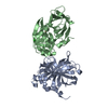
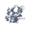
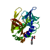
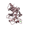
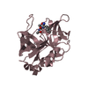
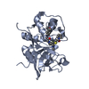
| Title | An episulfide cation (thiiranium ring) trapped in the active site of HAV 3C proteinase inactivated by peptide-based ketone inhibitors | ||||||
 Components Components |
| ||||||
 Keywords Keywords | HYDROLASE/HYDROLASE INHIBITOR / HEPATITIS A VIRUS / 3C PROTEASE / INHIBITOR DESIGN / METHYLKETONE / EPISULFIDE / PICORNAIN / HYDROLASE-HYDROLASE INHIBITOR COMPLEX | ||||||
| Function / homology |  Function and homology information Function and homology informationhost cell mitochondrial outer membrane / symbiont-mediated suppression of host cytoplasmic pattern recognition receptor signaling pathway via inhibition of MAVS activity / picornain 3C / T=pseudo3 icosahedral viral capsid / host cell cytoplasmic vesicle membrane / host multivesicular body / ribonucleoside triphosphate phosphatase activity / nucleoside-triphosphate phosphatase / channel activity / monoatomic ion transmembrane transport ...host cell mitochondrial outer membrane / symbiont-mediated suppression of host cytoplasmic pattern recognition receptor signaling pathway via inhibition of MAVS activity / picornain 3C / T=pseudo3 icosahedral viral capsid / host cell cytoplasmic vesicle membrane / host multivesicular body / ribonucleoside triphosphate phosphatase activity / nucleoside-triphosphate phosphatase / channel activity / monoatomic ion transmembrane transport / RNA helicase activity / RNA-directed RNA polymerase / cysteine-type endopeptidase activity / viral RNA genome replication / RNA-directed RNA polymerase activity / symbiont entry into host cell / virion attachment to host cell / DNA-templated transcription / structural molecule activity / proteolysis / RNA binding / ATP binding Similarity search - Function | ||||||
| Biological species |  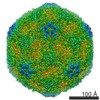 Hepatitis A virus Hepatitis A virus | ||||||
| Method |  X-RAY DIFFRACTION / SYNCHROTRON / MOLECULAR REPLACEMENT / Resolution: 1.35 Å X-RAY DIFFRACTION / SYNCHROTRON / MOLECULAR REPLACEMENT / Resolution: 1.35 Å | ||||||
 Authors Authors | Yin, J. / Cherney, M.M. / Bergmann, E.M. / James, M.N. | ||||||
 Citation Citation | Journal: J.Mol.Biol. / Year: 2006 Title: An Episulfide Cation (Thiiranium Ring) Trapped in the Active Site of HAV 3C Proteinase Inactivated by Peptide-based Ketone Inhibitors. Authors: Yin, J. / Cherney, M.M. / Bergmann, E.M. / Zhang, J. / Huitema, C. / Pettersson, H. / Eltis, L.D. / Vederas, J.C. / James, M.N. #1: Journal: J.Mol.Biol. / Year: 2005Title: Dual Modes of Modification of Hepatitis A Virus 3C Protease by a Serine-Derived beta-lactone: Selective Crystallization and formation of a functional catalytic triad in the active site Authors: Yin, J. / Cherney, M.M. / Bergmann, E.M. / Lall, M.S. / Jain, R.P. / Vederas, J.C. / James, M.N.G. #2: Journal: VIROLOGY / Year: 1999Title: Crystal Structure of an Inhibitor Complex of the 3C Proteinase from Hepatitis A Virus (HAV) and Implications for the Polyprotein Processing in HAV Authors: Bergmann, E.M. / Cherney, M.M. / Mckendrick, J. / Frormann, S. / Luo, C. / Malcolm, B.A. / Vederas, J.C. / James, M.N. #3: Journal: J.VIROL. / Year: 1997Title: The Refined Crystal Structure of the 3C Gene Product from Hepatitis A Virus: Specific Proteinase Activity and RNA Recognition Authors: Bergmann, E.M. / Mosimann, S.C. / Chernaia, M.M. / Malcolm, B.A. / James, M.N. | ||||||
| History |
|
- Structure visualization
| Structure viewer | Molecule: MolmilJmol/JSmol |
|---|
- Downloads & links
-Download
| PDBx/mmCIF format | 2hal.cif.gz | 69 KB | Display | PDBx/mmCIF format |
|---|---|---|---|---|
| PDB format | pdb2hal.ent.gz | 49.6 KB | Display | PDB format |
| PDBx/mmJSON format | 2hal.json.gz | Tree view | PDBx/mmJSON format | |
| Others |  Other downloads Other downloads |
-Validation report
| Arichive directory | https://data.pdbj.org/pub/pdb/validation_reports/ha/2halftp://data.pdbj.org/pub/pdb/validation_reports/ha/2hal | HTTPS FTP |
|---|
-Related structure data
| Related structure data | 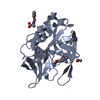 2h6mSC  2h9hC S: Starting model for refinement C: citing same article ( |
|---|---|
| Similar structure data |


-Links
 PDBj
PDBj

- Assembly
| Deposited unit | 
| ||||||||
|---|---|---|---|---|---|---|---|---|---|
| 1 |
| ||||||||
| Unit cell |
|
-Components
| #1: Protein | Mass: 23288.844 Da / Num. of mol.: 1 / Fragment: 3C proteinase, residues 1520-1731 / Mutation: C24S Source method: isolated from a genetically manipulated source Source: (gene. exp.) Hepatitis A virus / Genus: Hepatovirus / Gene: 3C / Plasmid: pHAV-3CEX / Production host:  References: UniProt: Q81090, UniProt: P08617*PLUS, picornain 3C |
|---|---|
| #2: Protein/peptide |   Type: Peptide-like / Class: Inhibitor / Mass: 598.706 Da / Num. of mol.: 1 / Source method: obtained synthetically Type: Peptide-like / Class: Inhibitor / Mass: 598.706 Da / Num. of mol.: 1 / Source method: obtained syntheticallyReferences: N-acetyl-L-leucyl-L-phenylalanyl-N-[(1S,2R)-1-(2-carboxyethyl)-3-fluoro-2-hydroxypropyl]-L-phenylalaninamide |
| #3: Chemical | ChemComp-BBL /   Mass: 223.225 Da / Num. of mol.: 1 / Source method: obtained synthetically / Formula: C11H13NO4 Mass: 223.225 Da / Num. of mol.: 1 / Source method: obtained synthetically / Formula: C11H13NO4 |
| #4: Water | ChemComp-HOH /  Mass: 18.015 Da / Num. of mol.: 435 / Source method: isolated from a natural source / Formula: H2O Mass: 18.015 Da / Num. of mol.: 435 / Source method: isolated from a natural source / Formula: H2O |
| Compound details | THE COORDINATES OF METHYLKETONE GLUTAMATE AND EPISULFIDE METHYLGLUTAMATE ARE ALTERNATE ...THE COORDINATE |
| Has protein modification | Y |
-Experimental details
-Experiment
| Experiment | Method: X-RAY DIFFRACTION / Number of used crystals: 1 |
|---|
- Sample preparation
| Crystal | Density Matthews: 2.14 Å3/Da / Density % sol: 42.51 % |
|---|---|
| Crystal grow | Temperature: 297 K / Method: vapor diffusion, hanging drop / pH: 7.5 Details: 2.5% PEG 8000, 1.5% Glycerol, 10mM Tris-HCl, pH 7.5, VAPOR DIFFUSION, HANGING DROP, temperature 297K |
-Data collection
| Diffraction | Mean temperature: 100 K |
|---|---|
| Diffraction source | Source: SYNCHROTRON / Site: ALS  / Beamline: 8.3.1 / Wavelength: 1.1159 Å / Beamline: 8.3.1 / Wavelength: 1.1159 Å |
| Detector | Type: ADSC QUANTUM 210 / Detector: CCD / Date: Jul 29, 2004 |
| Radiation | Monochromator: crystal / Protocol: SINGLE WAVELENGTH / Monochromatic (M) / Laue (L): M / Scattering type: x-ray |
| Radiation wavelength | Wavelength: 1.1159 Å / Relative weight: 1 |
| Reflection | Resolution: 1.35→40 Å / Num. all: 39555 / Num. obs: 38893 / % possible obs: 85.3 % / Observed criterion σ(F): 0 / Observed criterion σ(I): 0 / Rmerge(I) obs: 0.061 / Net I/σ(I): 13 |
| Reflection shell | Resolution: 1.35→1.4 Å / Rmerge(I) obs: 0.288 / % possible all: 42.4 |
- Processing
| Software |
| |||||||||||||||||||||||||||||||||||||||||||||||||||||||||||||||||||||||||||||||||||||||||||||||||||||||||
|---|---|---|---|---|---|---|---|---|---|---|---|---|---|---|---|---|---|---|---|---|---|---|---|---|---|---|---|---|---|---|---|---|---|---|---|---|---|---|---|---|---|---|---|---|---|---|---|---|---|---|---|---|---|---|---|---|---|---|---|---|---|---|---|---|---|---|---|---|---|---|---|---|---|---|---|---|---|---|---|---|---|---|---|---|---|---|---|---|---|---|---|---|---|---|---|---|---|---|---|---|---|---|---|---|---|---|
| Refinement | Method to determine structure: MOLECULAR REPLACEMENT Starting model: PDB ENTRY 2H6M Resolution: 1.35→20 Å / Cor.coef. Fo:Fc: 0.959 / Cor.coef. Fo:Fc free: 0.951 / SU B: 1.979 / SU ML: 0.038 / Cross valid method: THROUGHOUT / σ(F): 0 / ESU R: 0.092 / ESU R Free: 0.071 / Stereochemistry target values: MAXIMUM LIKELIHOOD / Details: HYDROGENS HAVE BEEN ADDED IN THE RIDING POSITIONS
| |||||||||||||||||||||||||||||||||||||||||||||||||||||||||||||||||||||||||||||||||||||||||||||||||||||||||
| Solvent computation | Ion probe radii: 0.8 Å / Shrinkage radii: 0.8 Å / VDW probe radii: 1.2 Å / Solvent model: MASK | |||||||||||||||||||||||||||||||||||||||||||||||||||||||||||||||||||||||||||||||||||||||||||||||||||||||||
| Displacement parameters | Biso mean: 17.008 Å2
| |||||||||||||||||||||||||||||||||||||||||||||||||||||||||||||||||||||||||||||||||||||||||||||||||||||||||
| Refinement step | Cycle: LAST / Resolution: 1.35→20 Å
| |||||||||||||||||||||||||||||||||||||||||||||||||||||||||||||||||||||||||||||||||||||||||||||||||||||||||
| Refine LS restraints |
| |||||||||||||||||||||||||||||||||||||||||||||||||||||||||||||||||||||||||||||||||||||||||||||||||||||||||
| LS refinement shell | Resolution: 1.35→1.385 Å / Total num. of bins used: 20
|
