 Movie
Movie Controller

[English] 日本語
 Yorodumi
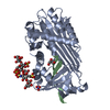
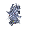
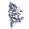
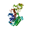
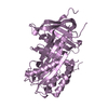
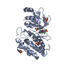
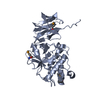
- PDB-2h4q: Crystal structure of a M-loop deletion variant of MENT in the cle... -
+ Open data

- Basic information
| Entry | Database: PDB / ID: 2h4q | ||||||
|---|---|---|---|---|---|---|---|
| Title | Crystal structure of a M-loop deletion variant of MENT in the cleaved conformation | ||||||
 Components Components | (Heterochromatin-associated protein MENT) x 2 | ||||||
 Keywords Keywords | HYDROLASE INHIBITOR / Serine protease inhibitor / Serpin | ||||||
| Function / homology |  Function and homology information Function and homology informationnucleate erythrocyte maturation / chromosome condensation / serine-type endopeptidase inhibitor activity / chromatin DNA binding / chromatin organization / chromatin / : / nucleoplasm Similarity search - Function | ||||||
| Biological species |  | ||||||
| Method |  X-RAY DIFFRACTION / MOLECULAR REPLACEMENT / Resolution: 2.1 Å X-RAY DIFFRACTION / MOLECULAR REPLACEMENT / Resolution: 2.1 Å | ||||||
 Authors Authors | Whisstock, J.C. / Buckle, A.M. / McGowan, S. / Irving, J.A. | ||||||
 Citation Citation | Journal: Embo J. / Year: 2006 Title: X-ray crystal structure of MENT: evidence for functional loop-sheet polymers in chromatin condensation. Authors: McGowan, S. / Buckle, A.M. / Irving, J.A. / Ong, P.C. / Bashtannyk-Puhalovich, T.A. / Kan, W.T. / Henderson, K.N. / Bulynko, Y.A. / Popova, E.Y. / Smith, A.I. / Bottomley, S.P. / Rossjohn, J. ...Authors: McGowan, S. / Buckle, A.M. / Irving, J.A. / Ong, P.C. / Bashtannyk-Puhalovich, T.A. / Kan, W.T. / Henderson, K.N. / Bulynko, Y.A. / Popova, E.Y. / Smith, A.I. / Bottomley, S.P. / Rossjohn, J. / Grigoryev, S.A. / Pike, R.N. / Whisstock, J.C. | ||||||
| History |
|
- Structure visualization
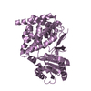
| Structure viewer | Molecule: MolmilJmol/JSmol |
|---|
- Downloads & links
-Download
| PDBx/mmCIF format | 2h4q.cif.gz | 100.5 KB | Display | PDBx/mmCIF format |
|---|---|---|---|---|
| PDB format | pdb2h4q.ent.gz | 74.8 KB | Display | PDB format |
| PDBx/mmJSON format | 2h4q.json.gz | Tree view | PDBx/mmJSON format | |
| Others |  Other downloads Other downloads |
-Validation report
| Arichive directory | https://data.pdbj.org/pub/pdb/validation_reports/h4/2h4qftp://data.pdbj.org/pub/pdb/validation_reports/h4/2h4q | HTTPS FTP |
|---|
-Related structure data
| Related structure data |  2dutC 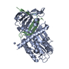 2h4pC 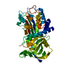 2h4rC  1qlpS C: citing same article ( S: Starting model for refinement |
|---|---|
| Similar structure data |


-Links
 PDBj
PDBj
- Assembly
| Deposited unit | 
| |||||||||||||||
|---|---|---|---|---|---|---|---|---|---|---|---|---|---|---|---|---|
| 1 |
| |||||||||||||||
| 2 | x 6
| |||||||||||||||
| Unit cell |
| |||||||||||||||
| Components on special symmetry positions |
|
-Components
| #1: Protein | Mass: 43953.160 Da / Num. of mol.: 1 / Fragment: residues 1-369 / Mutation: A361V, deletion mutant(residues 61-88) Source method: isolated from a genetically manipulated source Source: (gene. exp.)  |
|---|---|
| #2: Protein/peptide | Mass: 4186.003 Da / Num. of mol.: 1 / Fragment: residues 377-409 Source method: isolated from a genetically manipulated source Source: (gene. exp.) |
| #3: Water | ChemComp-HOH /  Mass: 18.015 Da / Num. of mol.: 350 / Source method: isolated from a natural source / Formula: H2O Mass: 18.015 Da / Num. of mol.: 350 / Source method: isolated from a natural source / Formula: H2O |
-Experimental details
-Experiment
| Experiment | Method: X-RAY DIFFRACTION / Number of used crystals: 1 |
|---|
- Sample preparation
| Crystal | Density Matthews: 3.97 Å3/Da / Density % sol: 68.99 % |
|---|---|
| Crystal grow | Temperature: 290 K / Method: vapor diffusion, hanging drop / pH: 6 Details: 0.1M sodium acetate, 12-15% PEG 8000, pH 5.5-6.5, pH 6.0, VAPOR DIFFUSION, HANGING DROP, temperature 290K |
-Data collection
| Diffraction | Mean temperature: 100 K | ||||||||||||||||||||||||||||||||||||||||||||||||||||||||||||||||||
|---|---|---|---|---|---|---|---|---|---|---|---|---|---|---|---|---|---|---|---|---|---|---|---|---|---|---|---|---|---|---|---|---|---|---|---|---|---|---|---|---|---|---|---|---|---|---|---|---|---|---|---|---|---|---|---|---|---|---|---|---|---|---|---|---|---|---|---|
| Diffraction source | Source: ROTATING ANODE / Type: ELLIOTT GX-13 / Wavelength: 1.5148 Å | ||||||||||||||||||||||||||||||||||||||||||||||||||||||||||||||||||
| Detector | Type: RIGAKU RAXIS IV / Detector: IMAGE PLATE / Date: Apr 7, 2003 / Details: OSMIC MIRRORS | ||||||||||||||||||||||||||||||||||||||||||||||||||||||||||||||||||
| Radiation | Protocol: SINGLE WAVELENGTH / Monochromatic (M) / Laue (L): M / Scattering type: x-ray | ||||||||||||||||||||||||||||||||||||||||||||||||||||||||||||||||||
| Radiation wavelength | Wavelength: 1.5148 Å / Relative weight: 1 | ||||||||||||||||||||||||||||||||||||||||||||||||||||||||||||||||||
| Reflection | Av σ(I) over netI: 14.4 / Number: 151605 / Rmerge(I) obs: 0.062 / Χ2: 1.25 / D res high: 2.1 Å / D res low: 50 Å / Num. obs: 44120 / % possible obs: 99.3 | ||||||||||||||||||||||||||||||||||||||||||||||||||||||||||||||||||
| Diffraction reflection shell |
| ||||||||||||||||||||||||||||||||||||||||||||||||||||||||||||||||||
| Reflection | Resolution: 2.1→105.4 Å / Num. all: 44451 / Num. obs: 44451 / % possible obs: 99.3 % / Observed criterion σ(F): 2 / Observed criterion σ(I): 2 / Redundancy: 3.4 % / Rmerge(I) obs: 0.062 / Net I/σ(I): 20.7 | ||||||||||||||||||||||||||||||||||||||||||||||||||||||||||||||||||
| Reflection shell | Resolution: 2.1→2.18 Å / Redundancy: 2.7 % / Rmerge(I) obs: 0.587 / Mean I/σ(I) obs: 2.4 / % possible all: 94.6 |
- Processing
| Software |
| |||||||||||||||||||||||||||||||||||||||||||||||||||||||||||||||||||||||||||||||||||||||||||||||||||||||||||||||||||||||||||||
|---|---|---|---|---|---|---|---|---|---|---|---|---|---|---|---|---|---|---|---|---|---|---|---|---|---|---|---|---|---|---|---|---|---|---|---|---|---|---|---|---|---|---|---|---|---|---|---|---|---|---|---|---|---|---|---|---|---|---|---|---|---|---|---|---|---|---|---|---|---|---|---|---|---|---|---|---|---|---|---|---|---|---|---|---|---|---|---|---|---|---|---|---|---|---|---|---|---|---|---|---|---|---|---|---|---|---|---|---|---|---|---|---|---|---|---|---|---|---|---|---|---|---|---|---|---|---|
| Refinement | Method to determine structure: MOLECULAR REPLACEMENT Starting model: PDB ENTRY 1QLP Resolution: 2.1→24.35 Å / Cor.coef. Fo:Fc: 0.959 / Cor.coef. Fo:Fc free: 0.95 / SU B: 7.437 / SU ML: 0.101 / Cross valid method: THROUGHOUT / σ(F): 0 / ESU R: 0.139 / ESU R Free: 0.13 / Stereochemistry target values: MAXIMUM LIKELIHOOD / Details: HYDROGENS HAVE BEEN ADDED IN THE RIDING POSITIONS
| |||||||||||||||||||||||||||||||||||||||||||||||||||||||||||||||||||||||||||||||||||||||||||||||||||||||||||||||||||||||||||||
| Solvent computation | Ion probe radii: 0.8 Å / Shrinkage radii: 0.8 Å / VDW probe radii: 1.2 Å / Solvent model: BABINET MODEL WITH MASK | |||||||||||||||||||||||||||||||||||||||||||||||||||||||||||||||||||||||||||||||||||||||||||||||||||||||||||||||||||||||||||||
| Displacement parameters | Biso mean: 32.052 Å2
| |||||||||||||||||||||||||||||||||||||||||||||||||||||||||||||||||||||||||||||||||||||||||||||||||||||||||||||||||||||||||||||
| Refinement step | Cycle: LAST / Resolution: 2.1→24.35 Å
| |||||||||||||||||||||||||||||||||||||||||||||||||||||||||||||||||||||||||||||||||||||||||||||||||||||||||||||||||||||||||||||
| Refine LS restraints |
| |||||||||||||||||||||||||||||||||||||||||||||||||||||||||||||||||||||||||||||||||||||||||||||||||||||||||||||||||||||||||||||
| LS refinement shell | Resolution: 2.1→2.155 Å / Total num. of bins used: 20
|
