 Movie
Movie Controller

[English] 日本語
 Yorodumi
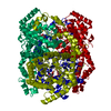
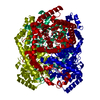
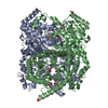
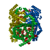
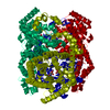
- PDB-2gyi: DESIGN, SYNTHESIS, AND CHARACTERIZATION OF A POTENT XYLOSE ISOMER... -
+ Open data

- Basic information
| Entry | Database: PDB / ID: 2gyi | |||||||||
|---|---|---|---|---|---|---|---|---|---|---|
| Title | DESIGN, SYNTHESIS, AND CHARACTERIZATION OF A POTENT XYLOSE ISOMERASE INHIBITOR, D-THREONOHYDROXAMIC ACID, AND HIGH-RESOLUTION X-RAY CRYSTALLOGRAPHIC STRUCTURE OF THE ENZYME-INHIBITOR COMPLEX | |||||||||
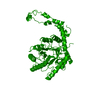
 Components Components | XYLOSE ISOMERASE | |||||||||
 Keywords Keywords | ISOMERASE(INTRAMOLECULAR OXIDOREDUCTASE) | |||||||||
| Function / homology |  Function and homology information Function and homology informationxylose isomerase / xylose isomerase activity / D-xylose metabolic process / magnesium ion binding / cytoplasm Similarity search - Function | |||||||||
| Biological species |  Streptomyces olivochromogenes (bacteria) Streptomyces olivochromogenes (bacteria) | |||||||||
| Method |  X-RAY DIFFRACTION / Resolution: 1.6 Å X-RAY DIFFRACTION / Resolution: 1.6 Å | |||||||||
 Authors Authors | Allen, K.N. / Lavie, A. / Petsko, G.A. / Ringe, D. | |||||||||
 Citation Citation | Journal: Biochemistry / Year: 1995 Title: Design, Synthesis, and Characterization of a Potent Xylose Isomerase Inhibitor, D-Threonohydroxamic Acid, and High-Resolution X-Ray Crystallographic Structure of the Enzyme-Inhibitor Complex Authors: Allen, K.N. / Lavie, A. / Petsko, G.A. / Ringe, D. #1: Journal: Biochemistry / Year: 1994Title: Isotopic Exchange Plus Substrate and Inhibition Kinetics of D-Xylose Isomerase Do not Support a Proton-Transfer Mechanism Authors: Allen, K.N. / Lavie, A. / Farber, G.K. / Glasfeld, A. / Petsko, G.A. / Ringe, D. #2: Journal: Biochemistry / Year: 1994Title: Role of the Divalent Metal Ion in Sugar Binding, Ring Opening, and Isomerization by D-Xylose Isomerase: Replacement of a Catalytic Metal by an Amino Acid Authors: Allen, K.N. / Lavie, A. / Glasfeld, A. / Tanada, T.N. / Gerrity, D.P. / Carlson, S.C. / Farber, G.K. / Petsko, G.A. / Ringe, D. #3: Journal: Biochemistry / Year: 1994Title: X-Ray Crystallographic Structures of D-Xylose Isomerase-Substrate Complexes Position the Substrate and Provide Evidence for Metal Movement During Catalysis Authors: Lavie, A. / Allen, K.N. / Petsko, G.A. / Ringe, D. | |||||||||
| History |
| |||||||||
| Remark 700 | SHEET THE SHEETS PRESENTED AS *S1* AND *S2* ON SHEET RECORDS BELOW ARE ACTUALLY EIGHT-STRANDED BETA- ...SHEET THE SHEETS PRESENTED AS *S1* AND *S2* ON SHEET RECORDS BELOW ARE ACTUALLY EIGHT-STRANDED BETA-BARRELS. THIS IS REPRESENTED BY NINE-STRANDED SHEETS IN WHICH THE FIRST AND LAST STRANDS OF EACH ARE IDENTICAL. |
- Structure visualization
| Structure viewer | Molecule: MolmilJmol/JSmol |
|---|
- Downloads & links
-Download
| PDBx/mmCIF format | 2gyi.cif.gz | 177.4 KB | Display | PDBx/mmCIF format |
|---|---|---|---|---|
| PDB format | pdb2gyi.ent.gz | 138.7 KB | Display | PDB format |
| PDBx/mmJSON format | 2gyi.json.gz | Tree view | PDBx/mmJSON format | |
| Others |  Other downloads Other downloads |
-Validation report
| Arichive directory | https://data.pdbj.org/pub/pdb/validation_reports/gy/2gyiftp://data.pdbj.org/pub/pdb/validation_reports/gy/2gyi | HTTPS FTP |
|---|
-Related structure data
| Similar structure data |
|---|

-Links
 PDBj
PDBj
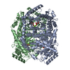
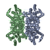
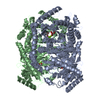
- Assembly
| Deposited unit | 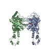
| ||||||||
|---|---|---|---|---|---|---|---|---|---|
| 1 | 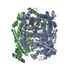
| ||||||||
| Unit cell |
| ||||||||
| Atom site foot note | 1: SER A 63 - ASP A 64 OMEGA = 247.14 PEPTIDE BOND DEVIATES SIGNIFICANTLY FROM TRANS CONFORMATION 2: CIS PROLINE - PRO A 186 / 3: CIS PROLINE - PRO B 186 | ||||||||
| Components on special symmetry positions |
| ||||||||
| Noncrystallographic symmetry (NCS) | NCS oper: (Code: given Matrix: (0.99927, 0.03829, -0.00045), Vector: Details | MTRIX THE TRANSFORMATIONS PRESENTED ON MTRIX RECORDS BELOW DESCRIBE NON-CRYSTALLOGRAPHIC RELATIONSHIPS AMONG THE VARIOUS DOMAINS IN THIS ENTRY. APPLYING THE APPROPRIATE MTRIX TRANSFORMATION TO THE RESIDUES LISTED FIRST WILL YIELD APPROXIMATE COORDINATES FOR THE RESIDUES LISTED SECOND. APPLIED TO TRANSFORMED TO MTRIX RESIDUES RESIDUES RMSD M1 A 2 .. A 386 B 2 .. B 386 1.200 | |
-Components
| #1: Protein | Mass: 42844.848 Da / Num. of mol.: 2 Source method: isolated from a genetically manipulated source Source: (gene. exp.) Streptomyces olivochromogenes (bacteria)References: UniProt: P15587, xylose isomerase #2: Chemical | ChemComp-MG /   Mass: 24.305 Da / Num. of mol.: 4 / Source method: obtained synthetically / Formula: Mg Mass: 24.305 Da / Num. of mol.: 4 / Source method: obtained synthetically / Formula: Mg#3: Chemical | 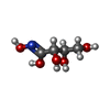  Mass: 151.118 Da / Num. of mol.: 2 / Source method: obtained synthetically / Formula: C4H9NO5 Mass: 151.118 Da / Num. of mol.: 2 / Source method: obtained synthetically / Formula: C4H9NO5#4: Water | ChemComp-HOH / |  Mass: 18.015 Da / Num. of mol.: 634 / Source method: isolated from a natural source / Formula: H2O Mass: 18.015 Da / Num. of mol.: 634 / Source method: isolated from a natural source / Formula: H2ONonpolymer details | THERE IS ONE D-THREONOHYDROXAMIC ACID MOLECULE BOUND IN THE LINEAR FORM IN THE ACTIVE SITE OF EACH ...THERE IS ONE D-THREONOHYD | |
|---|
-Experimental details
-Experiment
| Experiment | Method: X-RAY DIFFRACTION |
|---|
- Sample preparation
| Crystal | Density Matthews: 2.35 Å3/Da / Density % sol: 47.67 % | ||||||||||||||||||||||||||||||||||||
|---|---|---|---|---|---|---|---|---|---|---|---|---|---|---|---|---|---|---|---|---|---|---|---|---|---|---|---|---|---|---|---|---|---|---|---|---|---|
| Crystal grow | *PLUS pH: 7.5 / Method: vapor diffusion, sitting drop | ||||||||||||||||||||||||||||||||||||
| Components of the solutions | *PLUS
|
-Data collection
| Reflection | *PLUS Highest resolution: 1.6 Å / Lowest resolution: 44.72 Å / Num. obs: 83616 / Num. measured all: 190809 / Rmerge(I) obs: 0.051 |
|---|
- Processing
| Software |
| ||||||||||||||||||||||||||||||||||||||||||||||||||||||||||||
|---|---|---|---|---|---|---|---|---|---|---|---|---|---|---|---|---|---|---|---|---|---|---|---|---|---|---|---|---|---|---|---|---|---|---|---|---|---|---|---|---|---|---|---|---|---|---|---|---|---|---|---|---|---|---|---|---|---|---|---|---|---|
| Refinement | Resolution: 1.6→10 Å / σ(F): 0 /
| ||||||||||||||||||||||||||||||||||||||||||||||||||||||||||||
| Refinement step | Cycle: LAST / Resolution: 1.6→10 Å
| ||||||||||||||||||||||||||||||||||||||||||||||||||||||||||||
| Refine LS restraints |
| ||||||||||||||||||||||||||||||||||||||||||||||||||||||||||||
| Software | *PLUS Name: X-PLOR / Classification: refinement | ||||||||||||||||||||||||||||||||||||||||||||||||||||||||||||
| Refine LS restraints | *PLUS
|
