 Movie
Movie Controller

[English] 日本語
 Yorodumi
- PDB-2gxf: X-Ray Crystal Structure of Protein YybH from Bacillus subtilis. N... -
+ Open data

- Basic information
| Entry | Database: PDB / ID: 2gxf | ||||||
|---|---|---|---|---|---|---|---|
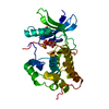
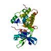
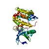
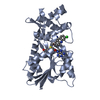
| Title | X-Ray Crystal Structure of Protein YybH from Bacillus subtilis. Northeast Structural Genomics Consortium Target SR506. | ||||||
 Components Components | Hypothetical protein yybH | ||||||
 Keywords Keywords | STRUCTURAL GENOMICS / UNKNOWN FUNCTION / alpha-beta protein. / PSI / Protein Structure Initiative / Northeast Structural Genomics Consortium / NESG | ||||||
| Function / homology | SnoaL-like domain / SnoaL-like domain / Nuclear Transport Factor 2; Chain: A, - #50 / NTF2-like domain superfamily / Nuclear Transport Factor 2; Chain: A, / Roll / Alpha Beta / Uncharacterized protein YybH Function and homology information Function and homology information | ||||||
| Biological species |  | ||||||
| Method |  X-RAY DIFFRACTION / SYNCHROTRON / MAD / Resolution: 3.1 Å X-RAY DIFFRACTION / SYNCHROTRON / MAD / Resolution: 3.1 Å | ||||||
 Authors Authors | Forouhar, F. / Abashidze, M. / Jayaraman, S. / Cunningham, K. / Fang, Y. / Ma, L.-C. / Xiao, R. / Acton, T.B. / Montelione, G.T. / Hunt, J.F. ...Forouhar, F. / Abashidze, M. / Jayaraman, S. / Cunningham, K. / Fang, Y. / Ma, L.-C. / Xiao, R. / Acton, T.B. / Montelione, G.T. / Hunt, J.F. / Tong, L. / Northeast Structural Genomics Consortium (NESG) | ||||||
 Citation Citation | Journal: TO BE PUBLISHED Title: Crystal Structure of the Hypothetical Protein YybH from Bacillus subtilis, Northeast Structural Genomics Target SR506 Authors: Forouhar, F. / Abashidze, M. / Jayaraman, S. / Cunningham, K. / Fang, Y. / Ma, L.-C. / Xiao, R. / Acton, T.B. / Montelione, G.T. / Hunt, J.F. / Tong, L. / Northeast Structural Genomics Consortium (NESG) | ||||||
| History |
|
- Structure visualization
| Structure viewer | Molecule: MolmilJmol/JSmol |
|---|
- Downloads & links
-Download
| PDBx/mmCIF format | 2gxf.cif.gz | 107.7 KB | Display | PDBx/mmCIF format |
|---|---|---|---|---|
| PDB format | pdb2gxf.ent.gz | 85.1 KB | Display | PDB format |
| PDBx/mmJSON format | 2gxf.json.gz | Tree view | PDBx/mmJSON format | |
| Others |  Other downloads Other downloads |
-Validation report
| Arichive directory | https://data.pdbj.org/pub/pdb/validation_reports/gx/2gxfftp://data.pdbj.org/pub/pdb/validation_reports/gx/2gxf | HTTPS FTP |
|---|
-Related structure data
| Similar structure data | |
|---|---|
| Other databases |


-Links
 PDBj
PDBj
- Assembly
| Deposited unit | 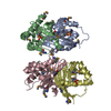
| ||||||||
|---|---|---|---|---|---|---|---|---|---|
| 1 | 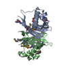
| ||||||||
| 2 | 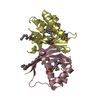
| ||||||||
| Unit cell |
|
-Components
| #1: Protein | Mass: 16408.705 Da / Num. of mol.: 4 Source method: isolated from a genetically manipulated source Source: (gene. exp.) #2: Chemical | ChemComp-MES /   Mass: 195.237 Da / Num. of mol.: 4 / Source method: obtained synthetically / Formula: C6H13NO4S / Comment: pH buffer*YM Mass: 195.237 Da / Num. of mol.: 4 / Source method: obtained synthetically / Formula: C6H13NO4S / Comment: pH buffer*YM#3: Water | ChemComp-HOH / |  Mass: 18.015 Da / Num. of mol.: 33 / Source method: isolated from a natural source / Formula: H2O Mass: 18.015 Da / Num. of mol.: 33 / Source method: isolated from a natural source / Formula: H2OHas protein modification | Y | |
|---|
-Experimental details
-Experiment
| Experiment | Method: X-RAY DIFFRACTION / Number of used crystals: 1 |
|---|
- Sample preparation
| Crystal | Density Matthews: 2.46 Å3/Da / Density % sol: 49.94 % Description: THE STRUCTURE FACTOR FILE CONTAINS FRIEDEL PAIRS. |
|---|---|
| Crystal grow | Temperature: 291 K / Method: vapor diffusion, hanging drop / pH: 6.15 Details: 100mM MES (pH 6.15), 22% PEG5kMME, 200mM ammonium sulfate, and 5mM DTT., VAPOR DIFFUSION, HANGING DROP, temperature 291K |
-Data collection
| Diffraction | Mean temperature: 100 K | ||||||||||||
|---|---|---|---|---|---|---|---|---|---|---|---|---|---|
| Diffraction source | Source: SYNCHROTRON / Site: NSLS  / Beamline: X4A / Wavelength: 0.97904, 0.97943, 0.96801 / Beamline: X4A / Wavelength: 0.97904, 0.97943, 0.96801 | ||||||||||||
| Detector | Type: ADSC QUANTUM 4 / Detector: CCD / Date: Feb 28, 2006 / Details: mirrors. | ||||||||||||
| Radiation | Monochromator: Si 111 CHANNEL / Protocol: MAD / Monochromatic (M) / Laue (L): M / Scattering type: x-ray | ||||||||||||
| Radiation wavelength |
| ||||||||||||
| Reflection | Resolution: 3.1→29.42 Å / Num. all: 22751 / Num. obs: 22660 / % possible obs: 99.6 % / Observed criterion σ(F): 0 / Observed criterion σ(I): 0 / Redundancy: 5 % / Biso Wilson estimate: 46.3 Å2 / Rmerge(I) obs: 0.112 / Rsym value: 0.081 / Net I/σ(I): 15.17 | ||||||||||||
| Reflection shell | Resolution: 3.1→3.21 Å / Redundancy: 5 % / Rmerge(I) obs: 0.585 / Mean I/σ(I) obs: 2.4 / Num. unique all: 2252 / Rsym value: 0.584 / % possible all: 100 |
- Processing
| Software |
| |||||||||||||||||||||||||
|---|---|---|---|---|---|---|---|---|---|---|---|---|---|---|---|---|---|---|---|---|---|---|---|---|---|---|
| Refinement | Method to determine structure: MAD / Resolution: 3.1→29.42 Å / Rfactor Rfree error: 0.007 / Data cutoff high absF: 411496.57 / Data cutoff low absF: 0 / Isotropic thermal model: OVERALL / Cross valid method: THROUGHOUT / σ(F): 2 / σ(I): 2 / Stereochemistry target values: Engh & Huber / Details: THE FRIEDEL PAIRS WERE USED FOR phasing.
| |||||||||||||||||||||||||
| Solvent computation | Solvent model: FLAT MODEL / Bsol: 10 Å2 / ksol: 0.266245 e/Å3 | |||||||||||||||||||||||||
| Displacement parameters | Biso mean: 52.4 Å2
| |||||||||||||||||||||||||
| Refine analyze |
| |||||||||||||||||||||||||
| Refinement step | Cycle: LAST / Resolution: 3.1→29.42 Å
| |||||||||||||||||||||||||
| Refine LS restraints |
| |||||||||||||||||||||||||
| LS refinement shell | Resolution: 3.1→3.29 Å / Rfactor Rfree error: 0.022 / Total num. of bins used: 6
|
