 Movie
Movie Controller


+ Open data

- Basic information
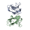
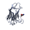
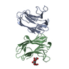
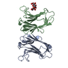
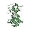
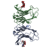
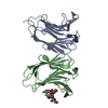
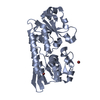
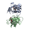
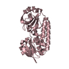
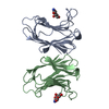
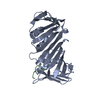
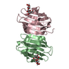
| Entry | Database: PDB / ID: 2gal | ||||||
|---|---|---|---|---|---|---|---|
| Title | CRYSTAL STRUCTURE OF HUMAN GALECTIN-7 IN COMPLEX WITH GALACTOSE | ||||||
 Components Components | GALECTIN-7 | ||||||
 Keywords Keywords | LECTIN / GALAPTIN / GALECTIN / CARBOHYDRATE BINDING | ||||||
| Function / homology |  Function and homology information Function and homology informationDifferentiation of Keratinocytes in Interfollicular Epidermis in Mammalian Skin / heterophilic cell-cell adhesion / carbohydrate binding / apoptotic process / : / extracellular exosome / nucleus / cytoplasm Similarity search - Function | ||||||
| Biological species |  Homo sapiens (human) Homo sapiens (human) | ||||||
| Method |  X-RAY DIFFRACTION / SYNCHROTRON / MOLECULAR REPLACEMENT / Resolution: 2 Å X-RAY DIFFRACTION / SYNCHROTRON / MOLECULAR REPLACEMENT / Resolution: 2 Å | ||||||
 Authors Authors | Leonidas, D.D. / Acharya, K.R. | ||||||
 Citation Citation | Journal: Biochemistry / Year: 1998 Title: Structural basis for the recognition of carbohydrates by human galectin-7. Authors: Leonidas, D.D. / Vatzaki, E.H. / Vorum, H. / Celis, J.E. / Madsen, P. / Acharya, K.R. | ||||||
| History |
|
- Structure visualization
| Structure viewer | Molecule: MolmilJmol/JSmol |
|---|
- Downloads & links
-Download
| PDBx/mmCIF format | 2gal.cif.gz | 63.9 KB | Display | PDBx/mmCIF format |
|---|---|---|---|---|
| PDB format | pdb2gal.ent.gz | 47.9 KB | Display | PDB format |
| PDBx/mmJSON format | 2gal.json.gz | Tree view | PDBx/mmJSON format | |
| Others |  Other downloads Other downloads |
-Validation report
| Arichive directory | https://data.pdbj.org/pub/pdb/validation_reports/ga/2galftp://data.pdbj.org/pub/pdb/validation_reports/ga/2gal | HTTPS FTP |
|---|
-Related structure data

-Links
 PDBj
PDBj

- Assembly
| Deposited unit | 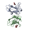
| ||||||||
|---|---|---|---|---|---|---|---|---|---|
| 1 |
| ||||||||
| Unit cell |
| ||||||||
| Noncrystallographic symmetry (NCS) | NCS oper: (Code: given Matrix: (-0.364826, -0.736594, 0.569501), Vector: |
-Components
| #1: Protein | Mass: 14965.850 Da / Num. of mol.: 2 Source method: isolated from a genetically manipulated source Source: (gene. exp.) Homo sapiens (human) / Cell line: BL21 / Plasmid: BL21 / Species (production host): Escherichia coli / Production host:  #2: Sugar |   Type: D-saccharide, beta linking / Mass: 180.156 Da / Num. of mol.: 2 Type: D-saccharide, beta linking / Mass: 180.156 Da / Num. of mol.: 2Source method: isolated from a genetically manipulated source Formula: C6H12O6 #3: Water | ChemComp-HOH / |  Mass: 18.015 Da / Num. of mol.: 76 / Source method: isolated from a natural source / Formula: H2O Mass: 18.015 Da / Num. of mol.: 76 / Source method: isolated from a natural source / Formula: H2O |
|---|
-Experimental details
-Experiment
| Experiment | Method: X-RAY DIFFRACTION / Number of used crystals: 1 |
|---|
- Sample preparation
| Crystal | Density Matthews: 2.2 Å3/Da / Density % sol: 43 % | ||||||||||||||||||||||||||||||||||||||||||||||||||||||||||||
|---|---|---|---|---|---|---|---|---|---|---|---|---|---|---|---|---|---|---|---|---|---|---|---|---|---|---|---|---|---|---|---|---|---|---|---|---|---|---|---|---|---|---|---|---|---|---|---|---|---|---|---|---|---|---|---|---|---|---|---|---|---|
| Crystal grow | Method: vapor diffusion, hanging drop / pH: 8.1 Details: CRYSTALS WERE GROWN USING THE HANGING DROP METHOD FROM DROPS CONTAINING 9 MG/ML PROTEIN AT PH 8.1 IN 50 MM SODIUM PHOSPHATE BUFFER, 0.3 M SODIUM CHLORIDE, 20 MM IMIDAZOLE, 8.5% PEG 3350 AND ...Details: CRYSTALS WERE GROWN USING THE HANGING DROP METHOD FROM DROPS CONTAINING 9 MG/ML PROTEIN AT PH 8.1 IN 50 MM SODIUM PHOSPHATE BUFFER, 0.3 M SODIUM CHLORIDE, 20 MM IMIDAZOLE, 8.5% PEG 3350 AND 25 MM GALACTOSE. DROPS WERE EQUILIBRATED AGAINST RESERVOIRS CONTAINING 50 MM SODIUM PHOSPHATE BUFFER, 0.3 M SODIUM CHLORIDE, 20 MM IMIDAZOLE, 17% PEG 3350 AND 50 MM GALACTOSE., vapor diffusion - hanging drop | ||||||||||||||||||||||||||||||||||||||||||||||||||||||||||||
| Crystal | *PLUS | ||||||||||||||||||||||||||||||||||||||||||||||||||||||||||||
| Crystal grow | *PLUS Temperature: 16 ℃ / Method: vapor diffusion, hanging drop | ||||||||||||||||||||||||||||||||||||||||||||||||||||||||||||
| Components of the solutions | *PLUS
|
-Data collection
| Diffraction | Mean temperature: 289 K |
|---|---|
| Diffraction source | Source: SYNCHROTRON / Site: EMBL/DESY, HAMBURG  / Beamline: X11 / Wavelength: 0.912 / Beamline: X11 / Wavelength: 0.912 |
| Detector | Type: MARRESEARCH / Detector: IMAGE PLATE / Date: Aug 1, 1997 / Details: MIRRORS |
| Radiation | Monochromatic (M) / Laue (L): M / Scattering type: x-ray |
| Radiation wavelength | Wavelength: 0.912 Å / Relative weight: 1 |
| Reflection | Resolution: 2→40 Å / Num. obs: 18155 / % possible obs: 98.2 % / Observed criterion σ(I): -3 / Redundancy: 9.5 % / Biso Wilson estimate: 8.4 Å2 / Rmerge(I) obs: 0.077 / Net I/σ(I): 9 |
| Reflection shell | Resolution: 2→2.1 Å / Redundancy: 4.5 % / Mean I/σ(I) obs: 6.6 / Rsym value: 0.384 / % possible all: 97.4 |
| Reflection | *PLUS Num. measured all: 172647 |
| Reflection shell | *PLUS % possible obs: 97.4 % |
- Processing
| Software |
| ||||||||||||||||||||||||||||||||||||||||||||||||||||||||||||||||||||||||||||||||
|---|---|---|---|---|---|---|---|---|---|---|---|---|---|---|---|---|---|---|---|---|---|---|---|---|---|---|---|---|---|---|---|---|---|---|---|---|---|---|---|---|---|---|---|---|---|---|---|---|---|---|---|---|---|---|---|---|---|---|---|---|---|---|---|---|---|---|---|---|---|---|---|---|---|---|---|---|---|---|---|---|---|
| Refinement | Method to determine structure: MOLECULAR REPLACEMENT Starting model: FREE GALECTIN-7 Resolution: 2→20 Å / Rfactor Rfree error: 0.008 / Data cutoff high absF: 10000000 / Data cutoff low absF: 0.001 / Isotropic thermal model: RESTRAINED / Cross valid method: THROUGHOUT / σ(F): 0 / Details: BULK SOLVENT MODEL USED
| ||||||||||||||||||||||||||||||||||||||||||||||||||||||||||||||||||||||||||||||||
| Displacement parameters | Biso mean: 26.2 Å2
| ||||||||||||||||||||||||||||||||||||||||||||||||||||||||||||||||||||||||||||||||
| Refine analyze |
| ||||||||||||||||||||||||||||||||||||||||||||||||||||||||||||||||||||||||||||||||
| Refinement step | Cycle: LAST / Resolution: 2→20 Å
| ||||||||||||||||||||||||||||||||||||||||||||||||||||||||||||||||||||||||||||||||
| Refine LS restraints |
| ||||||||||||||||||||||||||||||||||||||||||||||||||||||||||||||||||||||||||||||||
| LS refinement shell | Resolution: 2→2.13 Å / Rfactor Rfree error: 0.024 / Total num. of bins used: 6
| ||||||||||||||||||||||||||||||||||||||||||||||||||||||||||||||||||||||||||||||||
| Xplor file |
| ||||||||||||||||||||||||||||||||||||||||||||||||||||||||||||||||||||||||||||||||
| Software | *PLUS Name: X-PLOR / Version: 3.851 / Classification: refinement | ||||||||||||||||||||||||||||||||||||||||||||||||||||||||||||||||||||||||||||||||
| Refine LS restraints | *PLUS
|
