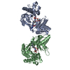
BIOMOLECULE: 1 THIS ENTRY CONTAINS THE CRYSTALLOGRAPHIC ASYMMETRIC UNIT WHICH CONSISTS OF 2 CHAIN(S) ...BIOMOLECULE: 1 THIS ENTRY CONTAINS THE CRYSTALLOGRAPHIC ASYMMETRIC UNIT WHICH CONSISTS OF 2 CHAIN(S). SEE REMARK 350 FOR INFORMATION ON GENERATING THE BIOLOGICAL MOLECULE(S). SIZE EXCLUSION CHROMATOGRAPHY WITH STATIC LIGHT SCATTERING SUPPORTS THE ASSIGNMENT OF A HEXAMER AS A BIOLOGICALLY SIGNIFICANT OLIGIMERIZATION STATE.
Resolution: 2.04→40.76 Å / Num. obs: 45685 / % possible obs: 99.7 % / Redundancy: 3.78 % / Biso Wilson estimate: 38.086 Å2 / Rmerge(I) obs: 0.067 / Net I/σ(I): 12.28
Reflection shell
Diffraction-ID: 1
Resolution (Å)
Highest resolution (Å)
Redundancy (%)
Rmerge(I) obs
Mean I/σ(I) obs
Num. measured obs
Num. unique obs
% possible all
2.04-2.11
3.39
0.437
2.93
13092
3925
96.9
2.11-2.2
0.303
4.7
17825
4658
95.4
2.2-2.3
0.242
5.8
16746
4366
96.7
2.3-2.42
0.181
7.5
17033
4434
97.5
2.42-2.57
0.139
9.2
17173
4468
98.3
2.57-2.77
0.102
11.7
17523
4545
98.5
2.77-3.04
0.078
14.4
16947
4397
99.3
3.04-3.48
0.056
18.1
17785
4630
99.6
3.48
0.048
21.9
17412
4589
99.8
-
Phasing
Phasing
Method: MAD
-
Processing
Software
Name
Version
Classification
NB
REFMAC
5.2.0005
refinement
XSCALE
datascaling
PDB_EXTRACT
1.701
dataextraction
XDS
datareduction
SHELX
phasing
autoSHARP
phasing
Refinement
Method to determine structure: MAD / Resolution: 2.04→39.01 Å / Cor.coef. Fo:Fc: 0.974 / Cor.coef. Fo:Fc free: 0.957 / SU B: 6.881 / SU ML: 0.093 / TLS residual ADP flag: LIKELY RESIDUAL / Cross valid method: THROUGHOUT / σ(F): 0 / ESU R: 0.135 / ESU R Free: 0.126 Stereochemistry target values: MAXIMUM LIKELIHOOD WITH PHASES Details: 1. HYDROGENS HAVE BEEN ADDED IN THE RIDING POSITIONS. 2. THERE IS ONE UNKNOWN LIGAND NEAR THE PUTATIVE ACTIVE SITE (CLOSE TO ASP156) FOR EACH MONOMER. 3. A MET-INHIBITION PROTOCOL WAS USED ...Details: 1. HYDROGENS HAVE BEEN ADDED IN THE RIDING POSITIONS. 2. THERE IS ONE UNKNOWN LIGAND NEAR THE PUTATIVE ACTIVE SITE (CLOSE TO ASP156) FOR EACH MONOMER. 3. A MET-INHIBITION PROTOCOL WAS USED FOR SELENOMETHIONINE INCORPORATION DURING PROTEIN EXPRESSION. THE OCCUPANCY OF THE SE ATOMS IN THE MSE RESIDUES WAS REDUCED TO 0.7 TO ACCOUNT FOR THE REDUCED SCATTERING POWER DUE TO PARTIAL S-MET INCORPORATION.
Rfactor
Num. reflection
% reflection
Selection details
Rfree
0.182
2287
5 %
RANDOM
Rwork
0.144
-
-
-
obs
0.146
45659
99.69 %
-
Solvent computation
Ion probe radii: 0.8 Å / Shrinkage radii: 0.8 Å / VDW probe radii: 1.2 Å / Solvent model: BABINET MODEL WITH MASK
Displacement parameters
Biso mean: 32.674 Å2
Baniso -1
Baniso -2
Baniso -3
1-
1.38 Å2
0.69 Å2
0 Å2
2-
-
1.38 Å2
0 Å2
3-
-
-
-2.07 Å2
Refinement step
Cycle: LAST / Resolution: 2.04→39.01 Å
Protein
Nucleic acid
Ligand
Solvent
Total
Num. atoms
4442
0
36
306
4784
Refine LS restraints
Refine-ID
Type
Dev ideal
Dev ideal target
Number
X-RAY DIFFRACTION
r_bond_refined_d
0.017
0.022
4616
X-RAY DIFFRACTION
r_bond_other_d
0.001
0.02
4453
X-RAY DIFFRACTION
r_angle_refined_deg
1.345
1.991
6226
X-RAY DIFFRACTION
r_angle_other_deg
0.767
3
10355
X-RAY DIFFRACTION
r_dihedral_angle_1_deg
5.894
5
601
X-RAY DIFFRACTION
r_dihedral_angle_2_deg
34.59
24.974
193
X-RAY DIFFRACTION
r_dihedral_angle_3_deg
13.124
15
894
X-RAY DIFFRACTION
r_dihedral_angle_4_deg
18.011
15
26
X-RAY DIFFRACTION
r_chiral_restr
0.08
0.2
731
X-RAY DIFFRACTION
r_gen_planes_refined
0.005
0.02
5075
X-RAY DIFFRACTION
r_gen_planes_other
0.001
0.02
879
X-RAY DIFFRACTION
r_nbd_refined
0.213
0.2
970
X-RAY DIFFRACTION
r_nbd_other
0.167
0.2
4473
X-RAY DIFFRACTION
r_nbtor_refined
0.176
0.2
2294
X-RAY DIFFRACTION
r_nbtor_other
0.083
0.2
2830
X-RAY DIFFRACTION
r_xyhbond_nbd_refined
0.168
0.2
244
X-RAY DIFFRACTION
r_symmetry_vdw_refined
0.241
0.2
11
X-RAY DIFFRACTION
r_symmetry_vdw_other
0.201
0.2
32
X-RAY DIFFRACTION
r_symmetry_hbond_refined
0.17
0.2
8
X-RAY DIFFRACTION
r_mcbond_it
2.219
3
2994
X-RAY DIFFRACTION
r_mcbond_other
0.526
3
1176
X-RAY DIFFRACTION
r_mcangle_it
3.045
5
4680
X-RAY DIFFRACTION
r_scbond_it
5.629
8
1809
X-RAY DIFFRACTION
r_scangle_it
8.029
11
1529
LS refinement shell
Resolution: 2.04→2.093 Å / Total num. of bins used: 20
Rfactor
Num. reflection
% reflection
Rfree
0.3
172
-
Rwork
0.212
3056
-
obs
-
3228
96.01 %
Refinement TLS params.
Method: refined / Refine-ID: X-RAY DIFFRACTION
ID
L11 (°2)
L12 (°2)
L13 (°2)
L22 (°2)
L23 (°2)
L33 (°2)
S11 (Å °)
S12 (Å °)
S13 (Å °)
S21 (Å °)
S22 (Å °)
S23 (Å °)
S31 (Å °)
S32 (Å °)
S33 (Å °)
T11 (Å2)
T12 (Å2)
T13 (Å2)
T22 (Å2)
T23 (Å2)
T33 (Å2)
Origin x (Å)
Origin y (Å)
Origin z (Å)
1
0.7186
-0.6668
0.7054
0.9778
-0.5611
1.1821
0.1022
0.0041
-0.0258
0.0098
-0.0557
-0.0036
-0.0166
-0.12
-0.0465
-0.0096
0.0271
-0.0073
-0.0403
0.0132
-0.0306
39.508
68.967
31.365
2
2.3908
-0.0216
-1.2021
1.1114
-0.148
3.8965
-0.039
0.1916
-0.2059
0.133
0.0217
0.1877
0.3051
-0.6089
0.0174
-0.0575
0.0053
0.0237
-0.0272
-0.0108
-0.0383
15.088
69.206
37.088
3
1.0392
-1.4212
0.7785
2.2646
-0.569
1.3489
0.1225
0.0626
-0.0035
-0.087
-0.1565
-0.0662
0.2181
0.0795
0.034
0.0555
0.0088
-0.0235
-0.0938
0.0034
-0.0429
49.479
57.3
35.423
4
0.7067
0.5461
-0.1572
2.0885
0.4482
3.2475
-0.0275
0.0039
0.0396
-0.0837
-0.0608
-0.0611
0.0012
0.0509
0.0883
-0.0956
0.0653
-0.0179
-0.1155
0.0072
-0.0385
54.532
39.493
18.249
Refinement TLS group
Refine-ID: X-RAY DIFFRACTION / Selection: ALL
ID
Refine TLS-ID
Auth asym-ID
Label asym-ID
Auth seq-ID
Label seq-ID
1
1
A
A
0 - 67
12 - 79
2
2
A
A
68 - 284
80 - 296
3
3
B
B
2 - 67
14 - 79
4
4
B
B
68 - 287
80 - 299
+
About Yorodumi
-
News
-
Feb 9, 2022. New format data for meta-information of EMDB entries
New format data for meta-information of EMDB entries
Version 3 of the EMDB header file is now the official format.
The previous official version 1.9 will be removed from the archive.
In the structure databanks used in Yorodumi, some data are registered as the other names, "COVID-19 virus" and "2019-nCoV". Here are the details of the virus and the list of structure data.
Jan 31, 2019. EMDB accession codes are about to change! (news from PDBe EMDB page)
EMDB accession codes are about to change! (news from PDBe EMDB page)
The allocation of 4 digits for EMDB accession codes will soon come to an end. Whilst these codes will remain in use, new EMDB accession codes will include an additional digit and will expand incrementally as the available range of codes is exhausted. The current 4-digit format prefixed with “EMD-” (i.e. EMD-XXXX) will advance to a 5-digit format (i.e. EMD-XXXXX), and so on. It is currently estimated that the 4-digit codes will be depleted around Spring 2019, at which point the 5-digit format will come into force.
The EM Navigator/Yorodumi systems omit the EMD- prefix.
Related info.:Q: What is EMD? / ID/Accession-code notation in Yorodumi/EM Navigator
Yorodumi is a browser for structure data from EMDB, PDB, SASBDB, etc.
This page is also the successor to EM Navigator detail page, and also detail information page/front-end page for Omokage search.
The word "yorodu" (or yorozu) is an old Japanese word meaning "ten thousand". "mi" (miru) is to see.
Related info.:EMDB / PDB / SASBDB / Comparison of 3 databanks / Yorodumi Search / Aug 31, 2016. New EM Navigator & Yorodumi / Yorodumi Papers / Jmol/JSmol / Function and homology information / Changes in new EM Navigator and Yorodumi
 Movie
Movie Controller

 Yorodumi
 Open data
 Basic information
 Components
 Keywords
 Function and homology information
 Pyrococcus horikoshii (archaea)
 X-RAY DIFFRACTION /
  Authors
 Citation
 Structure visualization
 Downloads & links
 Other downloads
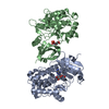


 PDBj
PDBj

 Assembly


 Mass: 62.068 Da / Num. of mol.: 5 / Source method: obtained synthetically / Formula: C2H6O2
 Mass: 18.015 Da / Num. of mol.: 306 / Source method: isolated from a natural source / Formula: H2O
 Sample preparation
 / Beamline: 8.2.2 / Wavelength: 1.0000, 0.9796, 0.9797
 Processing