 Movie
Movie Controller

[English] 日本語
 Yorodumi
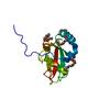
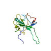
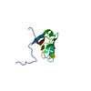
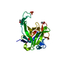
- PDB-2fvt: NMR Structure of the Rpa2829 protein from Rhodopseudomonas palust... -
+ Open data

- Basic information
| Entry | Database: PDB / ID: 2fvt | ||||||
|---|---|---|---|---|---|---|---|
| Title | NMR Structure of the Rpa2829 protein from Rhodopseudomonas palustris: Northeast Structural Genomics Target RpR43 | ||||||
 Components Components | conserved hypothetical protein | ||||||
 Keywords Keywords | STRUCTURAL GENOMICS / UNKNOWN FUNCTION / MTH938-like fold / PSI / Protein Structure Initiative / Northeast Structural Genomics Consortium / NESG | ||||||
| Function / homology | Hypothetical Protein Mth938; Chain: A, / MTH938-like / NDUFAF3/Mth938 domain-containing protein / MTH938-like superfamily / Protein of unknown function (DUF498/DUF598) / 3-Layer(aba) Sandwich / Alpha Beta / : / Mth938-like domain-containing protein Function and homology information Function and homology information | ||||||
| Biological species |  Rhodopseudomonas palustris (phototrophic) Rhodopseudomonas palustris (phototrophic) | ||||||
| Method | SOLUTION NMR / The initial structure was determined using automated structure determination (AutoStructure), refined manually. A final refinement used simultated annealing in explicit solvent. | ||||||
 Authors Authors | Cort, J.R. / Ho, C.K. / Cunningham, K. / Ma, L.C. / Conover, K. / Xiao, R. / Montelione, G.T. / Kennedy, M.A. / Northeast Structural Genomics Consortium (NESG) | ||||||
 Citation Citation | Journal: To be Published Title: NMR Structure of the Rpa2829 protein from Rhodopseudomonas palustris Authors: Cort, J.R. / Ho, C.K. / Cunningham, K. / Ma, L.C. / Conover, K. / Xiao, R. / Montelione, G.T. / Kennedy, M.A. | ||||||
| History |
| ||||||
| Remark 999 | Sequence According to authors,this is a sequencing error or an isoform resulting from a slight ...Sequence According to authors,this is a sequencing error or an isoform resulting from a slight difference in the sequenced strain and the cloning strain. |
- Structure visualization
| Structure viewer | Molecule: MolmilJmol/JSmol |
|---|
- Downloads & links
-Download
| PDBx/mmCIF format | 2fvt.cif.gz | 826.1 KB | Display | PDBx/mmCIF format |
|---|---|---|---|---|
| PDB format | pdb2fvt.ent.gz | 693 KB | Display | PDB format |
| PDBx/mmJSON format | 2fvt.json.gz | Tree view | PDBx/mmJSON format | |
| Others |  Other downloads Other downloads |
-Validation report
| Arichive directory | https://data.pdbj.org/pub/pdb/validation_reports/fv/2fvtftp://data.pdbj.org/pub/pdb/validation_reports/fv/2fvt | HTTPS FTP |
|---|
-Related structure data
| Similar structure data | |
|---|---|
| Other databases |

-Links
 PDBj
PDBj- Assembly
| Deposited unit | 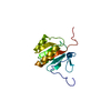
| |||||||||
|---|---|---|---|---|---|---|---|---|---|---|
| 1 |
| |||||||||
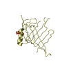
| NMR ensembles |
|
-Components
| #1: Protein | Mass: 15129.230 Da / Num. of mol.: 1 Source method: isolated from a genetically manipulated source Source: (gene. exp.) Rhodopseudomonas palustris (phototrophic)Gene: Rpa2829 / Plasmid: pET21 / Production host: |
|---|
-Experimental details
-Experiment
| Experiment | Method: SOLUTION NMR | ||||||||||||||||||||
|---|---|---|---|---|---|---|---|---|---|---|---|---|---|---|---|---|---|---|---|---|---|
| NMR experiment |
|
- Sample preparation
| Details |
| |||||||||||||||
|---|---|---|---|---|---|---|---|---|---|---|---|---|---|---|---|---|
| Sample conditions |
|
-NMR measurement
| Radiation | Protocol: SINGLE WAVELENGTH / Monochromatic (M) / Laue (L): M | |||||||||||||||||||||||||
|---|---|---|---|---|---|---|---|---|---|---|---|---|---|---|---|---|---|---|---|---|---|---|---|---|---|---|
| Radiation wavelength | Relative weight: 1 | |||||||||||||||||||||||||
| NMR spectrometer |
|
- Processing
| NMR software |
| ||||||||||||||||||||||||||||
|---|---|---|---|---|---|---|---|---|---|---|---|---|---|---|---|---|---|---|---|---|---|---|---|---|---|---|---|---|---|
| Refinement | Method: The initial structure was determined using automated structure determination (AutoStructure), refined manually. A final refinement used simultated annealing in explicit solvent. Software ordinal: 1 Details: THE STRUCTURES ARE BASED ON A TOTAL OF 1039 RESTRAINTS. SUMMARY OF EXPERIMENTAL CONSTRAINTS: RESTRAINING DISTANCE RESTRAINTS: TOTAL = 864; INTRA-RESIDUE [I=J] = 160; SEQUENTIAL [(I-J)=1] = ...Details: THE STRUCTURES ARE BASED ON A TOTAL OF 1039 RESTRAINTS. SUMMARY OF EXPERIMENTAL CONSTRAINTS: RESTRAINING DISTANCE RESTRAINTS: TOTAL = 864; INTRA-RESIDUE [I=J] = 160; SEQUENTIAL [(I-J)=1] = 225; MEDIUM RANGE [1<(I-J)<5] = 131; LONG RANGE [(I-J)>=5] = 348; HYDROGEN BOND RESTRAINTS = 72 (2 PER H-BOND); NUMBER OF RESTRAINING DISTANCE RESTRAINTS PER RESTRAINED RESIDUE = 8.2; DIHEDRAL-ANGLE RESTRAINTS = 103 (50 PHI, 52 PSI, 1 CHI-1); TOTAL NUMBER OF RESTRAINTS PER RESTRAINED RESIDUE = 8.9; NUMBER OF LONG RANGE NOE DISTANCE RESTRAINTS PER RESTRAINED RESIDUE = 3.0; NUMBER OF STRUCTURES COMPUTED = 30; NUMBER OF STRUCTURES USED = 20. AVERAGE DISTANCE VIOLATIONS >0.0001 ANG = 26.8+/-6; AVERAGE R.M.S. DISTANCE VIOLATION = 0.005 +/- 0.001 ANG; MAXIMUM NUMBER OF DISTANCE VIOLATIONS 39.MAXIMUM DISTANCE VIOLATION = 0.26 ANG; AVERAGE DIHEDRAL ANGLE VIOLATIONS: >0.0001 DEG = 3.3+/-1.3; MAX NUMBER OF DIHEDRAL ANGLE VIOLATIONS = 5; AVERAGE R.M.S. ANGLE VIOLATION = 0.42 +/- .01 DEG.; RMSD VALUES: BACKBONE ATOMS (N,C,C' RESIDUES 10-126) = 0.86 ANG, ALL HEAVY ATOMS = 1.52 ANG; BACKBONE ATOMS (N,C,C' RESIDUES 33-126) = 0.68 ANG, ALL HEAVY ATOMS = 1.1.38 ANG; BACKBONE ATOMS (N,C,C' RESIDUES 12-16,24-26,34-39,41-49,52-74,79-92,95-114,119-126) = 0.67 ANG,ALL HEAVY ATOMS = 1.19 ANG; PROCHECK (RESDIUES 10-126): MOST FAVORED REGIONS = 83.8%; ADDITIONAL ALLOWED REGIONS = 14.9%; GENEROUSLY ALLOWED REGIONS = 1.9%; DISALLOWED REGIONS = 0.4%. | ||||||||||||||||||||||||||||
| NMR representative | Selection criteria: few violations and low energy and good geometery | ||||||||||||||||||||||||||||
| NMR ensemble | Conformer selection criteria: structures with lowest energy and fewest restraint violations Conformers calculated total number: 30 / Conformers submitted total number: 20 |
 X-PLOR