 Movie
Movie Controller

[English] 日本語
 Yorodumi
- PDB-2fvo: Docking of the modified RF1 X-ray structure into the Low Resoluti... -
+ Open data

- Basic information
| Entry | Database: PDB / ID: 2fvo | ||||||
|---|---|---|---|---|---|---|---|
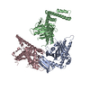
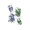
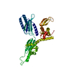
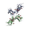
| Title | Docking of the modified RF1 X-ray structure into the Low Resolution Cryo-EM map of E.coli 70S Ribosome bound with RF1 | ||||||
 Components Components | Peptide chain release factor 1 | ||||||
 Keywords Keywords | TRANSLATION / RF1 ribosome cryo-EM | ||||||
| Function / homology |  Function and homology information Function and homology information | ||||||
| Biological species |   Thermotoga maritima (bacteria) Thermotoga maritima (bacteria) | ||||||
| Method | ELECTRON MICROSCOPY / single particle reconstruction / cryo EM / Resolution: 12.8 Å | ||||||
 Authors Authors | Rawat, U. / Gao, H. / Zavialov, A. / Gursky, R. / Ehrenberg, M. / Frank, J. | ||||||
 Citation Citation | Journal: J Mol Biol / Year: 2006 Title: Interactions of the release factor RF1 with the ribosome as revealed by cryo-EM. Authors: Urmila Rawat / Haixiao Gao / Andrey Zavialov / Richard Gursky / Måns Ehrenberg / Joachim Frank /  Abstract: In eubacteria, termination of translation is signaled by any one of the stop codons UAA, UAG, and UGA moving into the ribosomal A site. Two release factors, RF1 and RF2, recognize and bind to the ...In eubacteria, termination of translation is signaled by any one of the stop codons UAA, UAG, and UGA moving into the ribosomal A site. Two release factors, RF1 and RF2, recognize and bind to the stop codons with different affinities and trigger the hydrolysis of the ester bond that links the polypeptide with the P-site tRNA. Cryo-electron microscopy (cryo-EM) results obtained in this study show that ribosome-bound RF1 is in an open conformation, unlike the closed conformation observed in the crystal structure of the free factor, allowing its simultaneous access to both the decoding center and the peptidyl-transferase center. These results are similar to those obtained for RF2, but there is an important difference in how the factors bind to protein L11, which forms part of the GTPase-associated center of the large ribosomal subunit. The difference in the binding position, C-terminal domain for RF2 versus N-terminal domain for RF1, explains a body of L11 mutation studies that revealed differential effects on the activity of the two factors. Very recent data obtained with small-angle X-ray scattering now reveal that the solution structure of RF1 is open, as here seen on the ribosome by cryo-EM, and not closed, as seen in the crystal. #1: Journal: J Mol Biol / Year: 2004Title: Structural analyses of peptide release factor 1 from Thermotoga maritima reveal domain flexibility required for its interaction with the ribosome. Authors: Dong Hae Shin / Jeroen Brandsen / Jaru Jancarik / Hisao Yokota / Rosalind Kim / Sung-Hou Kim / Abstract: We have determined the crystal structure of peptide chain release factor 1 (RF1) from Thermotoga maritima (gi 4981173) at 2.65 Angstrom resolution by selenomethionine single-wavelength anomalous ...We have determined the crystal structure of peptide chain release factor 1 (RF1) from Thermotoga maritima (gi 4981173) at 2.65 Angstrom resolution by selenomethionine single-wavelength anomalous dispersion (SAD) techniques. RF1 is a protein that recognizes stop codons and promotes the release of a nascent polypeptide from tRNA on the ribosome. Selenomethionine-labeled RF1 crystallized in space group P2(1) with three monomers per asymmetric unit. It has approximate dimensions of 75 Angstrom x 70 Angstrom x 45 Angstrom and is composed of four domains. The overall fold of each RF1 domain shows almost the same topology with Escherichia coli RF2, except that the RF1 N-terminal domain is shorter and the C-terminal domain is longer than that of RF2. The N-terminal domain of RF1 indicates a rigid-body movement relative to that of RF2 with an angle of approximately 90 degrees. Including these features, RF1 has a tripeptide anticodon PVT motif instead of the SPF motif of RF2, which confers the specificity towards the stop codons. The analyses of three molecules in the asymmetric unit and comparison with RF2 revealed the presence of dynamic movement of domains I and III, which are anchored to the central domain by hinge loops. The crystal structure of RF1 elucidates the intrinsic property of this family of having large domain movements for proper function with the ribosome. | ||||||
| History |
|
- Structure visualization
| Movie |
Movie viewer |
|---|---|
| Structure viewer | Molecule: MolmilJmol/JSmol |
- Downloads & links
-Download
| PDBx/mmCIF format | 2fvo.cif.gz | 22.8 KB | Display | PDBx/mmCIF format |
|---|---|---|---|---|
| PDB format | pdb2fvo.ent.gz | 10.4 KB | Display | PDB format |
| PDBx/mmJSON format | 2fvo.json.gz | Tree view | PDBx/mmJSON format | |
| Others |  Other downloads Other downloads |
-Validation report
| Arichive directory | https://data.pdbj.org/pub/pdb/validation_reports/fv/2fvoftp://data.pdbj.org/pub/pdb/validation_reports/fv/2fvo | HTTPS FTP |
|---|
-Related structure data
| Related structure data | 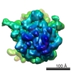 1184MC 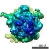 1185MC M: map data used to model this data C: citing same article ( |
|---|---|
| Similar structure data |


-Links
 PDBj
PDBj

- Assembly
| Deposited unit | 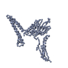
|
|---|---|
| 1 |
|
-Components
| #1: Protein | Mass: 38843.820 Da / Num. of mol.: 1 Source method: isolated from a genetically manipulated source Source: (gene. exp.) Thermotoga maritima (bacteria) / Gene: prfA / Production host: |
|---|
-Experimental details
-Experiment
| Experiment | Method: ELECTRON MICROSCOPY |
|---|---|
| EM experiment | Aggregation state: PARTICLE / 3D reconstruction method: single particle reconstruction |
- Sample preparation
| Component | Name: E.COLI 70S RIBOSOME - RF2 complex / Type: RIBOSOME |
|---|---|
| Buffer solution | Name: Polymix buffer / pH: 7.5 / Details: Polymix buffer |
| Specimen | Conc.: 32 mg/ml / Embedding applied: NO / Shadowing applied: NO / Staining applied: NO / Vitrification applied: YES |
| Specimen support | Details: quanti-foil grids coated with a thin carbon layer |
| Vitrification | Instrument: HOMEMADE PLUNGER / Cryogen name: ETHANE / Details: RAPID-FREEZING IN LIQUID Ethane |
- Electron microscopy imaging
| Experimental equipment |  Model: Tecnai F20 / Image courtesy: FEI Company |
|---|---|
| Microscopy | Model: FEI TECNAI F20 / Date: Dec 1, 2002 |
| Electron gun | Electron source:  FIELD EMISSION GUN / Accelerating voltage: 200 kV / Illumination mode: FLOOD BEAM FIELD EMISSION GUN / Accelerating voltage: 200 kV / Illumination mode: FLOOD BEAM |
| Electron lens | Mode: BRIGHT FIELD / Nominal magnification: 50000 X / Calibrated magnification: 49696 X / Nominal defocus max: 4000 nm / Nominal defocus min: 2000 nm / Cs: 2 mm |
| Specimen holder | Temperature: 93 K / Tilt angle max: 0 ° / Tilt angle min: 0 ° |
| Image recording | Electron dose: 20 e/Å2 / Film or detector model: KODAK SO-163 FILM |
- Processing
| EM software |
| ||||||||||||
|---|---|---|---|---|---|---|---|---|---|---|---|---|---|
| CTF correction | Details: CTF CORRECTION OF 3D-MAPS | ||||||||||||
| Symmetry | Point symmetry: C1 (asymmetric) | ||||||||||||
| 3D reconstruction | Method: Single particle / Resolution: 12.8 Å / Resolution method: FSC 0.5 CUT-OFF / Num. of particles: 24622 / Actual pixel size: 2.82 Å / Magnification calibration: TMV Details: Coordinates contain CA atoms only. ALl the image processing was done in SPIDER. 0.5 cutoff of FSC Symmetry type: POINT | ||||||||||||
| Atomic model building | Protocol: OTHER / Space: REAL / Details: METHOD--Manual | ||||||||||||
| Atomic model building | PDB-ID: 1RQ0 Accession code: 1RQ0 / Source name: PDB / Type: experimental model | ||||||||||||
| Refinement | Highest resolution: 12.8 Å / Details: Coordinates contain CA atoms only | ||||||||||||
| Refinement step | Cycle: LAST / Highest resolution: 12.8 Å
|
