 Movie
Movie Controller

[English] 日本語
 Yorodumi
- PDB-4es8: Crystal Structure of the adhesin domain of Epf from Streptococcus... -
+ Open data

- Basic information
| Entry | Database: PDB / ID: 4es8 | ||||||
|---|---|---|---|---|---|---|---|
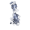
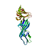
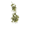
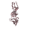
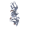
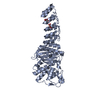
| Title | Crystal Structure of the adhesin domain of Epf from Streptococcus pyogenes in P212121 | ||||||
 Components Components | Epf | ||||||
 Keywords Keywords | CELL ADHESION / carbohydrate-binding module / fibronectin-like domain / adhesin / extracellular | ||||||
| Function / homology |  Function and homology information Function and homology informationJelly Rolls - #1240 / Immunoglobulin-like - #3580 / Domain of unknown function DUF1542 / Domain of Unknown Function (DUF1542) / LPXTG cell wall anchor motif / Gram-positive cocci surface proteins LPxTG motif profile. / LPXTG cell wall anchor domain / Jelly Rolls / Immunoglobulin-like / Sandwich / Mainly Beta Similarity search - Domain/homology | ||||||
| Biological species |  Streptococcus pyogenes (bacteria) Streptococcus pyogenes (bacteria) | ||||||
| Method |  X-RAY DIFFRACTION / SYNCHROTRON / MAD / Resolution: 1.58 Å X-RAY DIFFRACTION / SYNCHROTRON / MAD / Resolution: 1.58 Å | ||||||
 Authors Authors | Linke, C. / Siemens, N. / Kreikemeyer, B. / Baker, E.N. | ||||||
 Citation Citation | Journal: J.Biol.Chem. / Year: 2012 Title: The Extracellular Protein Factor Epf from Streptococcus pyogenes Is a Cell Surface Adhesin That Binds to Cells through an N-terminal Domain Containing a Carbohydrate-binding Module. Authors: Linke, C. / Siemens, N. / Oehmcke, S. / Radjainia, M. / Law, R.H. / Whisstock, J.C. / Baker, E.N. / Kreikemeyer, B. #1: Journal: To be PublishedTitle: Purification, crystallization and preliminary crystallographic analysis of the adhesion domain of Epf from Streptococcus pyogenes Authors: Linke, C. / Siemens, N. / Middleditch, M. / Kreikenmeyer, B. / Baker, E.N. | ||||||
| History |
|
- Structure visualization
| Structure viewer | Molecule: MolmilJmol/JSmol |
|---|
- Downloads & links
-Download
| PDBx/mmCIF format | 4es8.cif.gz | 160.5 KB | Display | PDBx/mmCIF format |
|---|---|---|---|---|
| PDB format | pdb4es8.ent.gz | 123.3 KB | Display | PDB format |
| PDBx/mmJSON format | 4es8.json.gz | Tree view | PDBx/mmJSON format | |
| Others |  Other downloads Other downloads |
-Validation report
| Arichive directory | https://data.pdbj.org/pub/pdb/validation_reports/es/4es8ftp://data.pdbj.org/pub/pdb/validation_reports/es/4es8 | HTTPS FTP |
|---|
-Related structure data


-Links
 PDBj
PDBj- Assembly
| Deposited unit | 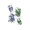
| ||||||||
|---|---|---|---|---|---|---|---|---|---|
| 1 | 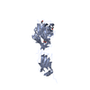
| ||||||||
| 2 | 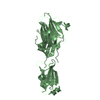
| ||||||||
| Unit cell |
|
-Components
-Protein , 1 types, 2 molecules AB
| #1: Protein | Mass: 35799.207 Da / Num. of mol.: 2 / Fragment: N-terminal domain Source method: isolated from a genetically manipulated source Source: (gene. exp.) Streptococcus pyogenes (bacteria) / Strain: M49 591 / Gene: SpyoM01000212 / Plasmid: pASK_IBA2 / Production host: |
|---|
-Non-polymers , 5 types, 983 molecules 


| #2: Chemical | ChemComp-GOL /  Mass: 92.094 Da / Num. of mol.: 1 / Source method: obtained synthetically / Formula: C3H8O3 Mass: 92.094 Da / Num. of mol.: 1 / Source method: obtained synthetically / Formula: C3H8O3 | ||||||
|---|---|---|---|---|---|---|---|
| #3: Chemical |  Mass: 59.044 Da / Num. of mol.: 2 / Source method: obtained synthetically / Formula: C2H3O2 Mass: 59.044 Da / Num. of mol.: 2 / Source method: obtained synthetically / Formula: C2H3O2#4: Chemical | ChemComp-NA / |  Mass: 22.990 Da / Num. of mol.: 1 / Source method: obtained synthetically / Formula: Na Mass: 22.990 Da / Num. of mol.: 1 / Source method: obtained synthetically / Formula: Na#5: Chemical | ChemComp-K / |  Mass: 39.098 Da / Num. of mol.: 1 / Source method: obtained synthetically / Formula: K Mass: 39.098 Da / Num. of mol.: 1 / Source method: obtained synthetically / Formula: K#6: Water | ChemComp-HOH / | Mass: 18.015 Da / Num. of mol.: 978 / Source method: isolated from a natural source / Formula: H2O |
-Experimental details
-Experiment
| Experiment | Method: X-RAY DIFFRACTION / Number of used crystals: 1 |
|---|
- Sample preparation
| Crystal | Density Matthews: 2.12 Å3/Da / Density % sol: 42.04 % |
|---|---|
| Crystal grow | Temperature: 291 K / Method: vapor diffusion, hanging drop / pH: 7.4 Details: 25 % (w/v) PEG3350, 400 mM K Acetate, pH 7.4, 0.02 MG/ML CHYMOTRYPSIN (Type VII, TLCK-treated, Sigma-Aldrich), temperature 291K, VAPOR DIFFUSION, HANGING DROP |
-Data collection
| Diffraction | Mean temperature: 100 K | ||||||||||||||||||||||||||||||||||||||||||||||||||||||||||||||||||||||||||||||||||||||||
|---|---|---|---|---|---|---|---|---|---|---|---|---|---|---|---|---|---|---|---|---|---|---|---|---|---|---|---|---|---|---|---|---|---|---|---|---|---|---|---|---|---|---|---|---|---|---|---|---|---|---|---|---|---|---|---|---|---|---|---|---|---|---|---|---|---|---|---|---|---|---|---|---|---|---|---|---|---|---|---|---|---|---|---|---|---|---|---|---|---|
| Diffraction source | Source: SYNCHROTRON / Site: Australian Synchrotron  / Beamline: MX2 / Wavelength: 0.979420, 0.95369, 0.97941 / Beamline: MX2 / Wavelength: 0.979420, 0.95369, 0.97941 | ||||||||||||||||||||||||||||||||||||||||||||||||||||||||||||||||||||||||||||||||||||||||
| Detector | Type: ADSC QUANTUM 315r / Detector: CCD / Date: Mar 11, 2001 | ||||||||||||||||||||||||||||||||||||||||||||||||||||||||||||||||||||||||||||||||||||||||
| Radiation | Protocol: MAD / Monochromatic (M) / Laue (L): M / Scattering type: x-ray | ||||||||||||||||||||||||||||||||||||||||||||||||||||||||||||||||||||||||||||||||||||||||
| Radiation wavelength |
| ||||||||||||||||||||||||||||||||||||||||||||||||||||||||||||||||||||||||||||||||||||||||
| Reflection | Resolution: 1.579→19.803 Å / Num. all: 83319 / Num. obs: 83319 / % possible obs: 98.7 % / Redundancy: 20.4 % / Rsym value: 0.07 / Net I/σ(I): 35.1 | ||||||||||||||||||||||||||||||||||||||||||||||||||||||||||||||||||||||||||||||||||||||||
| Reflection shell | Diffraction-ID: 1
|
- Processing
| Software |
| ||||||||||||||||||||||||||||||||||||||||||||||||||||||||||||||||||||||||||||||||||||||||||
|---|---|---|---|---|---|---|---|---|---|---|---|---|---|---|---|---|---|---|---|---|---|---|---|---|---|---|---|---|---|---|---|---|---|---|---|---|---|---|---|---|---|---|---|---|---|---|---|---|---|---|---|---|---|---|---|---|---|---|---|---|---|---|---|---|---|---|---|---|---|---|---|---|---|---|---|---|---|---|---|---|---|---|---|---|---|---|---|---|---|---|---|
| Refinement | Method to determine structure: MAD / Resolution: 1.58→19.8 Å / Cor.coef. Fo:Fc: 0.9615 / Cor.coef. Fo:Fc free: 0.9497 / Occupancy max: 1 / Occupancy min: 0.25 / Cross valid method: THROUGHOUT / σ(F): 0
| ||||||||||||||||||||||||||||||||||||||||||||||||||||||||||||||||||||||||||||||||||||||||||
| Displacement parameters | Biso max: 106.55 Å2 / Biso mean: 17.1438 Å2 / Biso min: 3.44 Å2
| ||||||||||||||||||||||||||||||||||||||||||||||||||||||||||||||||||||||||||||||||||||||||||
| Refine analyze | Luzzati coordinate error obs: 0.139 Å | ||||||||||||||||||||||||||||||||||||||||||||||||||||||||||||||||||||||||||||||||||||||||||
| Refinement step | Cycle: LAST / Resolution: 1.58→19.8 Å
| ||||||||||||||||||||||||||||||||||||||||||||||||||||||||||||||||||||||||||||||||||||||||||
| Refine LS restraints |
| ||||||||||||||||||||||||||||||||||||||||||||||||||||||||||||||||||||||||||||||||||||||||||
| LS refinement shell | Resolution: 1.58→1.62 Å / Total num. of bins used: 20
|
