 Movie
Movie Controller

[English] 日本語
 Yorodumi
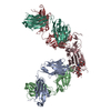
- PDB-2fjh: Structure of the B20-4 Fab, a phage derived Fab fragment, in comp... -
+ Open data

- Basic information
| Entry | Database: PDB / ID: 2fjh | ||||||
|---|---|---|---|---|---|---|---|
| Title | Structure of the B20-4 Fab, a phage derived Fab fragment, in complex with VEGF | ||||||
 Components Components |
| ||||||
 Keywords Keywords | HORMONE/GROWTH FACTOR/IMMUNE SYSTEM / VEGF / Fab / Protein Fab complex / Cystine knot / HORMONE-GROWTH FACTOR-IMMUNE SYSTEM COMPLEX | ||||||
| Function / homology |  Function and homology information Function and homology informationSignaling by VEGF / basophil chemotaxis / positive regulation of endothelial cell chemotaxis by VEGF-activated vascular endothelial growth factor receptor signaling pathway / cellular stress response to acid chemical / : / VEGF ligand-receptor interactions / vascular endothelial growth factor receptor 1 binding / negative regulation of adherens junction organization / primitive erythrocyte differentiation / positive regulation of mast cell chemotaxis ...Signaling by VEGF / basophil chemotaxis / positive regulation of endothelial cell chemotaxis by VEGF-activated vascular endothelial growth factor receptor signaling pathway / cellular stress response to acid chemical / : / VEGF ligand-receptor interactions / vascular endothelial growth factor receptor 1 binding / negative regulation of adherens junction organization / primitive erythrocyte differentiation / positive regulation of mast cell chemotaxis / negative regulation of establishment of endothelial barrier / vascular endothelial growth factor receptor binding / post-embryonic camera-type eye development / lymph vessel morphogenesis / negative regulation of blood-brain barrier permeability / VEGF-activated neuropilin signaling pathway / positive regulation of cell proliferation by VEGF-activated platelet derived growth factor receptor signaling pathway / eye photoreceptor cell development / motor neuron migration / coronary vein morphogenesis / cardiac vascular smooth muscle cell development / VEGF binds to VEGFR leading to receptor dimerization / vascular endothelial growth factor receptor-2 signaling pathway / positive regulation of axon extension involved in axon guidance / endothelial cell chemotaxis / positive regulation of protein localization to early endosome / positive regulation of trophoblast cell migration / surfactant homeostasis / positive regulation of protein autophosphorylation / camera-type eye morphogenesis / vascular wound healing / induction of positive chemotaxis / retinal ganglion cell axon guidance / dopaminergic neuron differentiation / positive regulation of epithelial tube formation / transmembrane receptor protein tyrosine kinase activator activity / neuropilin binding / positive regulation of blood vessel endothelial cell proliferation involved in sprouting angiogenesis / positive regulation of branching involved in ureteric bud morphogenesis / tube formation / coronary artery morphogenesis / negative regulation of cell-cell adhesion mediated by cadherin / positive regulation of leukocyte migration / cell migration involved in sprouting angiogenesis / vascular endothelial growth factor receptor 2 binding / positive regulation of vascular permeability / commissural neuron axon guidance / positive regulation of vascular endothelial growth factor signaling pathway / artery morphogenesis / cardiac muscle cell development / branching involved in blood vessel morphogenesis / positive regulation of blood vessel branching / sprouting angiogenesis / platelet-derived growth factor receptor binding / extracellular matrix binding / positive regulation of positive chemotaxis / Regulation of gene expression by Hypoxia-inducible Factor / positive regulation of neuroblast proliferation / positive regulation of endothelial cell chemotaxis / positive regulation of cell migration involved in sprouting angiogenesis / positive regulation of DNA biosynthetic process / negative regulation of epithelial to mesenchymal transition / vascular endothelial growth factor signaling pathway / mammary gland alveolus development / positive chemotaxis / positive regulation of sprouting angiogenesis / outflow tract morphogenesis / chemoattractant activity / mesoderm development / macrophage differentiation / fibronectin binding / monocyte differentiation / positive regulation of cell division / cellular response to vascular endothelial growth factor stimulus / vasculogenesis / lung development / positive regulation of receptor internalization / heart morphogenesis / positive regulation of focal adhesion assembly / vascular endothelial growth factor receptor signaling pathway / positive regulation of blood vessel endothelial cell migration / ovarian follicle development / cell maturation / lactation / kidney development / TFAP2 (AP-2) family regulates transcription of growth factors and their receptors / positive regulation of endothelial cell proliferation / epithelial cell differentiation / positive regulation of endothelial cell migration / negative regulation of miRNA transcription / positive regulation of cell adhesion / positive regulation of epithelial cell proliferation / secretory granule / platelet alpha granule lumen / cytokine activity / VEGFR2 mediated cell proliferation / growth factor activity / adherens junction / positive regulation of protein-containing complex assembly / positive regulation of protein phosphorylation Similarity search - Function | ||||||
| Biological species |  Homo sapiens (human) Homo sapiens (human) | ||||||
| Method |  X-RAY DIFFRACTION / SYNCHROTRON / MOLECULAR REPLACEMENT / Resolution: 3.1 Å X-RAY DIFFRACTION / SYNCHROTRON / MOLECULAR REPLACEMENT / Resolution: 3.1 Å | ||||||
 Authors Authors | Wiesmann, C. | ||||||
 Citation Citation | Journal: J.Biol.Chem. / Year: 2006 Title: Structure-function studies of two synthetic anti-vascular endothelial growth factor Fabs and comparison with the Avastin Fab. Authors: Fuh, G. / Wu, P. / Liang, W.C. / Ultsch, M. / Lee, C.V. / Moffat, B. / Wiesmann, C. | ||||||
| History |
| ||||||
| Remark 999 | SEQUENCE The Fab fragment of an antibody was derived using phage display. Therefore there is no ...SEQUENCE The Fab fragment of an antibody was derived using phage display. Therefore there is no match for the deposited FAB sequences in any sequence database. |
- Structure visualization
| Structure viewer | Molecule: MolmilJmol/JSmol |
|---|
- Downloads & links
-Download
| PDBx/mmCIF format | 2fjh.cif.gz | 210 KB | Display | PDBx/mmCIF format |
|---|---|---|---|---|
| PDB format | pdb2fjh.ent.gz | 170.2 KB | Display | PDB format |
| PDBx/mmJSON format | 2fjh.json.gz | Tree view | PDBx/mmJSON format | |
| Others |  Other downloads Other downloads |
-Validation report
| Arichive directory | https://data.pdbj.org/pub/pdb/validation_reports/fj/2fjhftp://data.pdbj.org/pub/pdb/validation_reports/fj/2fjh | HTTPS FTP |
|---|
-Related structure data
| Related structure data |  2fjfSC 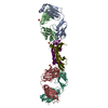 2fjgC 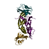 1fltS S: Starting model for refinement C: citing same article ( |
|---|---|
| Similar structure data |


-Links
 PDBj
PDBj


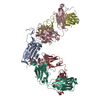
- Assembly
| Deposited unit | 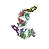
| ||||||||
|---|---|---|---|---|---|---|---|---|---|
| 1 | 
| ||||||||
| 2 | 
| ||||||||
| Unit cell |
| ||||||||
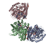
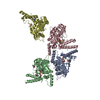
| Details | The crystallographic two-fold axis generates two NCS related complexes comprising a homodimer of VEGF bound to 2 Fab moleucles |
-Components
| #1: Protein | Mass: 11948.680 Da / Num. of mol.: 2 / Fragment: Receptor binding domain of VEGF (residues 34-135) Source method: isolated from a genetically manipulated source Source: (gene. exp.) Homo sapiens (human) / Plasmid: pB2105 / Production host:  #2: Antibody | Mass: 23253.875 Da / Num. of mol.: 2 Source method: isolated from a genetically manipulated source Source: (gene. exp.) Homo sapiens (human) / Plasmid: PW0357-4 / Production host: #3: Antibody | Mass: 24198.023 Da / Num. of mol.: 2 Source method: isolated from a genetically manipulated source Source: (gene. exp.) Homo sapiens (human) / Plasmid: PW0357-4 / Production host: Has protein modification | Y | |
|---|
-Experimental details
-Experiment
| Experiment | Method: X-RAY DIFFRACTION / Number of used crystals: 1 |
|---|
- Sample preparation
| Crystal | Density Matthews: 5.046684 Å3/Da / Density % sol: 75.627563 % |
|---|---|
| Crystal grow | Temperature: 292 K / Method: vapor diffusion, sitting drop / pH: 4.6 Details: 0.1 M Sodium acetate, 1.6 M Ammonium sulfate, pH 4.6, VAPOR DIFFUSION, SITTING DROP, temperature 292K |
-Data collection
| Diffraction | Mean temperature: 100 K |
|---|---|
| Diffraction source | Source: SYNCHROTRON / Site: ALS  / Beamline: 8.3.1 / Wavelength: 1 Å / Beamline: 8.3.1 / Wavelength: 1 Å |
| Detector | Type: ADSC QUANTUM 4 / Detector: CCD / Date: Mar 14, 2004 |
| Radiation | Protocol: SINGLE WAVELENGTH / Monochromatic (M) / Laue (L): M / Scattering type: x-ray |
| Radiation wavelength | Wavelength: 1 Å / Relative weight: 1 |
| Reflection | Resolution: 3.1→50 Å / Num. all: 42840 / Num. obs: 42755 / % possible obs: 99.8 % / Observed criterion σ(F): 0 / Observed criterion σ(I): 0 |
| Reflection shell | Resolution: 3.1→3.21 Å / % possible all: 100 |
- Processing
| Software |
| |||||||||||||||||||||||||||||||||||||||||||||||||||||||||||||||||||||||||||||||||||||||||||||||||||||||||||||||||||||||||||||||||||||||||||||||||||||||||||||||||||||||||||||||||||||||||||||||||||||||||||||||||||||||||||||||||||||||||||||||||||||||||||||||||||||||||||||||||||
|---|---|---|---|---|---|---|---|---|---|---|---|---|---|---|---|---|---|---|---|---|---|---|---|---|---|---|---|---|---|---|---|---|---|---|---|---|---|---|---|---|---|---|---|---|---|---|---|---|---|---|---|---|---|---|---|---|---|---|---|---|---|---|---|---|---|---|---|---|---|---|---|---|---|---|---|---|---|---|---|---|---|---|---|---|---|---|---|---|---|---|---|---|---|---|---|---|---|---|---|---|---|---|---|---|---|---|---|---|---|---|---|---|---|---|---|---|---|---|---|---|---|---|---|---|---|---|---|---|---|---|---|---|---|---|---|---|---|---|---|---|---|---|---|---|---|---|---|---|---|---|---|---|---|---|---|---|---|---|---|---|---|---|---|---|---|---|---|---|---|---|---|---|---|---|---|---|---|---|---|---|---|---|---|---|---|---|---|---|---|---|---|---|---|---|---|---|---|---|---|---|---|---|---|---|---|---|---|---|---|---|---|---|---|---|---|---|---|---|---|---|---|---|---|---|---|---|---|---|---|---|---|---|---|---|---|---|---|---|---|---|---|---|---|---|---|---|---|---|---|---|---|---|---|---|---|---|---|---|---|---|---|---|---|---|---|---|---|---|---|---|---|---|---|---|---|---|
| Refinement | Method to determine structure: MOLECULAR REPLACEMENT Starting model: Search model for VEGF was based on 1FLT, search model for Fab was based on 2FJF. Resolution: 3.1→30 Å / Cor.coef. Fo:Fc: 0.949 / Cor.coef. Fo:Fc free: 0.927 / SU B: 15.736 / SU ML: 0.267 / TLS residual ADP flag: LIKELY RESIDUAL / Cross valid method: THROUGHOUT / σ(F): 0 / ESU R: 0.657 / ESU R Free: 0.343 / Stereochemistry target values: MAXIMUM LIKELIHOOD / Details: HYDROGENS HAVE BEEN ADDED IN THE RIDING POSITIONS
| |||||||||||||||||||||||||||||||||||||||||||||||||||||||||||||||||||||||||||||||||||||||||||||||||||||||||||||||||||||||||||||||||||||||||||||||||||||||||||||||||||||||||||||||||||||||||||||||||||||||||||||||||||||||||||||||||||||||||||||||||||||||||||||||||||||||||||||||||||
| Solvent computation | Ion probe radii: 0.8 Å / Shrinkage radii: 0.8 Å / VDW probe radii: 1.4 Å / Solvent model: MASK | |||||||||||||||||||||||||||||||||||||||||||||||||||||||||||||||||||||||||||||||||||||||||||||||||||||||||||||||||||||||||||||||||||||||||||||||||||||||||||||||||||||||||||||||||||||||||||||||||||||||||||||||||||||||||||||||||||||||||||||||||||||||||||||||||||||||||||||||||||
| Displacement parameters | Biso mean: 55.043 Å2
| |||||||||||||||||||||||||||||||||||||||||||||||||||||||||||||||||||||||||||||||||||||||||||||||||||||||||||||||||||||||||||||||||||||||||||||||||||||||||||||||||||||||||||||||||||||||||||||||||||||||||||||||||||||||||||||||||||||||||||||||||||||||||||||||||||||||||||||||||||
| Refinement step | Cycle: LAST / Resolution: 3.1→30 Å
| |||||||||||||||||||||||||||||||||||||||||||||||||||||||||||||||||||||||||||||||||||||||||||||||||||||||||||||||||||||||||||||||||||||||||||||||||||||||||||||||||||||||||||||||||||||||||||||||||||||||||||||||||||||||||||||||||||||||||||||||||||||||||||||||||||||||||||||||||||
| Refine LS restraints |
| |||||||||||||||||||||||||||||||||||||||||||||||||||||||||||||||||||||||||||||||||||||||||||||||||||||||||||||||||||||||||||||||||||||||||||||||||||||||||||||||||||||||||||||||||||||||||||||||||||||||||||||||||||||||||||||||||||||||||||||||||||||||||||||||||||||||||||||||||||
| LS refinement shell | Resolution: 3.1→3.163 Å / Total num. of bins used: 25 /
| |||||||||||||||||||||||||||||||||||||||||||||||||||||||||||||||||||||||||||||||||||||||||||||||||||||||||||||||||||||||||||||||||||||||||||||||||||||||||||||||||||||||||||||||||||||||||||||||||||||||||||||||||||||||||||||||||||||||||||||||||||||||||||||||||||||||||||||||||||
| Refinement TLS params. | Method: refined / Refine-ID: X-RAY DIFFRACTION
| |||||||||||||||||||||||||||||||||||||||||||||||||||||||||||||||||||||||||||||||||||||||||||||||||||||||||||||||||||||||||||||||||||||||||||||||||||||||||||||||||||||||||||||||||||||||||||||||||||||||||||||||||||||||||||||||||||||||||||||||||||||||||||||||||||||||||||||||||||
| Refinement TLS group |
|
