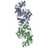
| Deposited unit | A: gelsolin
B: gelsolin
hetero molecules
| Theoretical mass | Number of molelcules |
|---|
| Total (without water) | 162,858 | 4 |
|---|
| Polymers | 161,844 | 2 |
|---|
| Non-polymers | 1,014 | 2 |
|---|
| Water | 901 | 50 |
|---|
|
|---|
| 1 |
- Idetical with deposited unit
- defined by author&software
| Type | Name | Symmetry operation | Number |
|---|
| identity operation | 1_555 | x,y,z | 1 |
| Buried area | 4060 Å2 |
|---|
| ΔGint | -13 kcal/mol |
|---|
| Surface area | 60540 Å2 |
|---|
| Method | PISA |
|---|
|
|---|
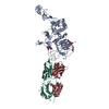
| 2 | A: gelsolin
B: gelsolin
hetero molecules
A: gelsolin
B: gelsolin
hetero molecules
A: gelsolin
B: gelsolin
hetero molecules
A: gelsolin
B: gelsolin
hetero molecules
| Theoretical mass | Number of molelcules |
|---|
| Total (without water) | 651,432 | 16 |
|---|
| Polymers | 647,375 | 8 |
|---|
| Non-polymers | 4,057 | 8 |
|---|
| Water | 144 | 8 |
|---|
| Type | Name | Symmetry operation | Number |
|---|
| identity operation | 1_555 | x,y,z | 1 | | crystal symmetry operation | 2_565 | -x,-y+1,z | 1 | | crystal symmetry operation | 3_555 | -y+1/2,x+1/2,z | 1 | | crystal symmetry operation | 4_455 | y-1/2,-x+1/2,z | 1 |
| Buried area | 31200 Å2 |
|---|
| ΔGint | -115 kcal/mol |
|---|
| Surface area | 227200 Å2 |
|---|
| Method | PISA |
|---|
|
|---|
| Unit cell | | Length a, b, c (Å) | 167.344, 167.344, 149.883 |
|---|
| Angle α, β, γ (deg.) | 90.00, 90.00, 90.00 |
|---|
| Int Tables number | 90 |
|---|
| Space group name H-M | P4212 |
|---|
|
|---|
| Noncrystallographic symmetry (NCS) | NCS domain: | ID | Ens-ID | Details (eV) |
|---|
| 1 | 1 | A| 2 | 1 | B| 1 | 2 | A| 2 | 2 | B| 1 | 3 | A| 2 | 3 | B| 1 | 4 | A| 2 | 4 | B| 1 | 5 | A| 2 | 5 | B| 1 | 6 | A| 2 | 6 | B| 1 | 7 | A| 2 | 7 | B| 1 | 8 | A| 2 | 8 | B| 1 | 9 | A| 2 | 9 | B | | | | | | | | | | | | | | | | | |
NCS domain segments: Component-ID: 1 / Refine code: 1 | Dom-ID | Ens-ID | Beg auth comp-ID | Beg label comp-ID | End auth comp-ID | End label comp-ID | Auth asym-ID | Label asym-ID | Auth seq-ID | Label seq-ID |
|---|
| 1 | 1 | VALVALLYSLYSAA| 27 - 135 | 3 - 111 | | 2 | 1 | VALVALLYSLYSBB| 27 - 135 | 3 - 111 | | 1 | 2 | SERSERGLYGLYAA| 136 - 248 | 112 - 224 | | 2 | 2 | SERSERGLYGLYBB| 136 - 248 | 112 - 224 | | 1 | 3 | PROPROLYSLYSAA| 249 - 367 | 225 - 343 | | 2 | 3 | PROPROLYSLYSBB| 249 - 367 | 225 - 343 | | 1 | 4 | ASNASNVALVALAA| 368 - 393 | 344 - 369 | | 2 | 4 | ASNASNVALVALBB| 368 - 393 | 344 - 369 | | 1 | 5 | PROPROGLYGLYAA| 394 - 513 | 370 - 489 | | 2 | 5 | PROPROGLYGLYBB| 394 - 513 | 370 - 489 | | 1 | 6 | GLYGLYGLYGLYAA| 514 - 619 | 490 - 595 | | 2 | 6 | GLYGLYGLYGLYBB| 514 - 619 | 490 - 595 | | 1 | 7 | GLYGLYLEULEUA| A | | | | | | | | | | | | | | | | | | | | | | | | | | | | | | | | | | | | | | | | | | | | | | | | | | | | | | | | | | | | | | | | | | | | | | | | | | | | | |
|
|---|
 Movie
Movie Controller


 Open data
 Basic information
 Components
 Keywords
 Function and homology information
 X-RAY DIFFRACTION /
  Authors
 Citation
 Structure visualization
 Downloads & links
 Other downloads
 PDBj
PDBj
 Assembly