Mass: 18.015 Da / Num. of mol.: 1522 / Source method: isolated from a natural source / Formula: H2O
-
Details
Sequence details
THE CONSTRUCT WAS EXPRESSED WITH A PURIFICATION TAG MGSDKIHHHHHHENLYFQG. THE TAG WAS REMOVED WITH ...THE CONSTRUCT WAS EXPRESSED WITH A PURIFICATION TAG MGSDKIHHHHHHENLYFQG. THE TAG WAS REMOVED WITH TEV PROTEASE LEAVING ONLY A GLYCINE (0) FOLLOWED BY THE TARGET SEQUENCE.
-
Experimental details
-
Experiment
Experiment
Method: X-RAY DIFFRACTION / Number of used crystals: 1
-
Sample preparation
Crystal
Density Matthews: 2.38 Å3/Da / Density % sol: 48.3 %
Crystal grow
Temperature: 277 K / pH: 8.36 Details: 53.80% 2-methyl-2,4-pentanediol, 0.1M TRIS pH 8.36, NANODROP, VAPOR DIFFUSION, SITTING DROP, temperature 277K
Resolution: 1.8→29.699 Å / Num. obs: 138522 / % possible obs: 94.4 % / Observed criterion σ(I): -3 / Biso Wilson estimate: 16.78 Å2
Reflection shell
Resolution: 1.8→1.86 Å / Rmerge(I) obs: 0.371 / Mean I/σ(I) obs: 1.9 / % possible all: 95.3
-
Phasing
Phasing
Method: MAD
-
Processing
Software
Name
Version
Classification
NB
SOLVE
phasing
REFMAC
5.5.0110
refinement
XSCALE
datascaling
PDB_EXTRACT
3.1
dataextraction
qfit
0.9
modelbuilding
XDS
datareduction
Refinement
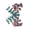
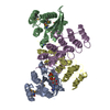
Method to determine structure: MAD / Resolution: 1.8→29.7 Å / Cor.coef. Fo:Fc: 0.956 / Cor.coef. Fo:Fc free: 0.938 / Occupancy max: 1 / Occupancy min: 0.21 / SU B: 4.781 / SU ML: 0.079 / ESU R: 0.124 / ESU R Free: 0.116 Stereochemistry target values: MAXIMUM LIKELIHOOD WITH PHASES Details: 1. HYDROGENS HAVE BEEN ADDED IN THE RIDING POSITIONS. 2. A MET-INHIBITION PROTOCOL WAS USED FOR SELENOMETHIONINE INCORPORATION DURING PROTEIN EXPRESSION. THE OCCUPANCY OF THE SE ATOMS IN THE ...Details: 1. HYDROGENS HAVE BEEN ADDED IN THE RIDING POSITIONS. 2. A MET-INHIBITION PROTOCOL WAS USED FOR SELENOMETHIONINE INCORPORATION DURING PROTEIN EXPRESSION. THE OCCUPANCY OF THE SE ATOMS IN THE MSE RESIDUES WAS REDUCED TO 0.75 FOR THE REDUCED SCATTERING POWER DUE TO PARTIAL S-MET INCORPORATION. 3. ATOM RECORD CONTAINS SUM OF TLS AND RESIDUAL B FACTORS. ANISOU RECORD CONTAINS SUM OF TLS AND RESIDUAL U FACTORS. 4. WATERS WERE EXCLUDED FROM AUTOMATIC TLS ASSIGNMENT. 5.ELECTRON DENSITY INDICATES THAT LYS 207 ON THE FOUR SUBUNITS IN THE ASYMMETRIC UNIT ARE COVALENTLY ATTACHED TO PYRIDOXAL-5'- PHOSPHATE; THERFORE THESE RESIDUES WERE MODELED AS 2-LYSINE (3- HYDROXY-2-METHYL-5-PHOSPHONOOXYMETHYL-PYRIDIN-4-YLMETHANE)(LLP). 6. 2-METHYL-2,4-PENTANEDIOL (MPD,MRD), CHLORIDE (CL), AND SODIUM IONS FROM THE CRYSTALLIZATION SOLUTION WERE MODELED INTO THE STRUCTURE. 7. THE PROGARM QFIT THAT AUTOMATICALLY IDENTIFIES AND MODELS DISCRETE HETEROGENEITY IN ELECTRON DENSITY MAPS WITH A A CONVEX OPTIMIZATION ALGORITHM WAS IMPLEMENTED DURING REFINEMENT TO MODEL ALTERNATE CONFORMATIONS.
In the structure databanks used in Yorodumi, some data are registered as the other names, "COVID-19 virus" and "2019-nCoV". Here are the details of the virus and the list of structure data.
Jan 31, 2019. EMDB accession codes are about to change! (news from PDBe EMDB page)
EMDB accession codes are about to change! (news from PDBe EMDB page)
The allocation of 4 digits for EMDB accession codes will soon come to an end. Whilst these codes will remain in use, new EMDB accession codes will include an additional digit and will expand incrementally as the available range of codes is exhausted. The current 4-digit format prefixed with “EMD-” (i.e. EMD-XXXX) will advance to a 5-digit format (i.e. EMD-XXXXX), and so on. It is currently estimated that the 4-digit codes will be depleted around Spring 2019, at which point the 5-digit format will come into force.
The EM Navigator/Yorodumi systems omit the EMD- prefix.
Related info.:Q: What is EMD? / ID/Accession-code notation in Yorodumi/EM Navigator
Yorodumi is a browser for structure data from EMDB, PDB, SASBDB, etc.
This page is also the successor to EM Navigator detail page, and also detail information page/front-end page for Omokage search.
The word "yorodu" (or yorozu) is an old Japanese word meaning "ten thousand". "mi" (miru) is to see.
Related info.:EMDB / PDB / SASBDB / Comparison of 3 databanks / Yorodumi Search / Aug 31, 2016. New EM Navigator & Yorodumi / Yorodumi Papers / Jmol/JSmol / Function and homology information / Changes in new EM Navigator and Yorodumi
 Movie
Movie Controller

 Yorodumi
 Open data
 Basic information
 Components
 Keywords
 Function and homology information
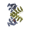
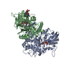
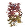
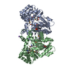
 Listeria monocytogenes (bacteria)
 X-RAY DIFFRACTION /
  Authors
 Citation
 Structure visualization
 Downloads & links
 Other downloads


 PDBj
PDBj Assembly
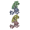


 Mass: 118.174 Da / Num. of mol.: 6 / Source method: obtained synthetically / Formula: C6H14O2 / Comment: precipitant*YM
 Mass: 22.990 Da / Num. of mol.: 2 / Source method: obtained synthetically / Formula: Na
 Mass: 35.453 Da / Num. of mol.: 1 / Source method: obtained synthetically / Formula: Cl
 Mass: 118.174 Da / Num. of mol.: 1 / Source method: obtained synthetically / Formula: C6H14O2 / Comment: precipitant*YM
 Sample preparation
 / Beamline: BL9-2 / Wavelength: 0.96109,0.97915,0.97936
 Processing