 Movie
Movie Controller


+ Open data

- Basic information
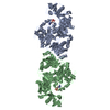
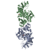
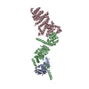
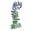
| Entry | Database: PDB / ID: 6att | |||||||||
|---|---|---|---|---|---|---|---|---|---|---|
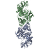
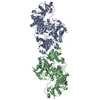
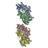
| Title | 39S Fab bound to HER2 ecd | |||||||||
 Components Components |
| |||||||||
 Keywords Keywords | IMMUNE SYSTEM / Antibody / fragment antigen binding / HER2 / receptor | |||||||||
| Function / homology |  Function and homology information Function and homology informationnegative regulation of immature T cell proliferation in thymus / ERBB3:ERBB2 complex / ERBB2-ERBB4 signaling pathway / immature T cell proliferation in thymus / GRB7 events in ERBB2 signaling / RNA polymerase I core binding / semaphorin receptor complex / Developmental Lineage of Mammary Stem Cells / ErbB-3 class receptor binding / Sema4D induced cell migration and growth-cone collapse ...negative regulation of immature T cell proliferation in thymus / ERBB3:ERBB2 complex / ERBB2-ERBB4 signaling pathway / immature T cell proliferation in thymus / GRB7 events in ERBB2 signaling / RNA polymerase I core binding / semaphorin receptor complex / Developmental Lineage of Mammary Stem Cells / ErbB-3 class receptor binding / Sema4D induced cell migration and growth-cone collapse / motor neuron axon guidance / regulation of microtubule-based process / PLCG1 events in ERBB2 signaling / enzyme-linked receptor protein signaling pathway / ERBB2 Activates PTK6 Signaling / ERBB2-EGFR signaling pathway / ERBB2-ERBB3 signaling pathway / neurotransmitter receptor localization to postsynaptic specialization membrane / Drug-mediated inhibition of ERBB2 signaling / Resistance of ERBB2 KD mutants to trastuzumab / Resistance of ERBB2 KD mutants to sapitinib / Resistance of ERBB2 KD mutants to tesevatinib / Resistance of ERBB2 KD mutants to neratinib / Resistance of ERBB2 KD mutants to osimertinib / Resistance of ERBB2 KD mutants to afatinib / Resistance of ERBB2 KD mutants to AEE788 / Resistance of ERBB2 KD mutants to lapatinib / Drug resistance in ERBB2 TMD/JMD mutants / positive regulation of MAP kinase activity / neuromuscular junction development / positive regulation of Rho protein signal transduction / positive regulation of transcription by RNA polymerase I / ERBB2 Regulates Cell Motility / Developmental Lineage of Mammary Gland Myoepithelial Cells / oligodendrocyte differentiation / semaphorin-plexin signaling pathway / PI3K events in ERBB2 signaling / regulation of angiogenesis / positive regulation of protein targeting to membrane / regulation of ERK1 and ERK2 cascade / Schwann cell development / coreceptor activity / Signaling by ERBB2 / TFAP2 (AP-2) family regulates transcription of growth factors and their receptors / peptidyl-tyrosine phosphorylation / myelination / transmembrane receptor protein tyrosine kinase activity / GRB2 events in ERBB2 signaling / positive regulation of cell adhesion / cell surface receptor protein tyrosine kinase signaling pathway / SHC1 events in ERBB2 signaling / basal plasma membrane / cellular response to epidermal growth factor stimulus / Constitutive Signaling by Overexpressed ERBB2 / positive regulation of epithelial cell proliferation / Downregulation of ERBB2:ERBB3 signaling / positive regulation of translation / neuromuscular junction / wound healing / phosphatidylinositol 3-kinase/protein kinase B signal transduction / Signaling by ERBB2 TMD/JMD mutants / receptor protein-tyrosine kinase / Signaling by ERBB2 ECD mutants / Signaling by ERBB2 KD Mutants / receptor tyrosine kinase binding / cellular response to growth factor stimulus / epidermal growth factor receptor signaling pathway / ruffle membrane / Downregulation of ERBB2 signaling / Constitutive Signaling by Aberrant PI3K in Cancer / neuron differentiation / transmembrane signaling receptor activity / PIP3 activates AKT signaling / myelin sheath / heart development / PI5P, PP2A and IER3 Regulate PI3K/AKT Signaling / positive regulation of cell growth / RAF/MAP kinase cascade / protein tyrosine kinase activity / presynaptic membrane / basolateral plasma membrane / protein phosphorylation / early endosome / positive regulation of MAPK cascade / cell surface receptor signaling pathway / signaling receptor complex / cell population proliferation / endosome membrane / apical plasma membrane / intracellular signal transduction / protein heterodimerization activity / signaling receptor binding / negative regulation of apoptotic process / perinuclear region of cytoplasm / signal transduction / nucleoplasm / ATP binding / membrane / identical protein binding / nucleus Similarity search - Function | |||||||||
| Biological species |  Homo sapiens (human) Homo sapiens (human) | |||||||||
| Method |  X-RAY DIFFRACTION / SYNCHROTRON / MOLECULAR REPLACEMENT / Resolution: 3.77 Å X-RAY DIFFRACTION / SYNCHROTRON / MOLECULAR REPLACEMENT / Resolution: 3.77 Å | |||||||||
 Authors Authors | Oganesyan, V.Y. / Dall'Acqua, W.F. | |||||||||
 Citation Citation | Journal: J. Biol. Chem. / Year: 2018 Title: Structural insights into the mechanism of action of a biparatopic anti-HER2 antibody. Authors: Oganesyan, V. / Peng, L. / Bee, J.S. / Li, J. / Perry, S.R. / Comer, F. / Xu, L. / Cook, K. / Senthil, K. / Clarke, L. / Rosenthal, K. / Gao, C. / Damschroder, M. / Wu, H. / Dall'Acqua, W. | |||||||||
| History |
|
- Structure visualization
| Structure viewer | Molecule: MolmilJmol/JSmol |
|---|
- Downloads & links
-Download
| PDBx/mmCIF format | 6att.cif.gz | 210.2 KB | Display | PDBx/mmCIF format |
|---|---|---|---|---|
| PDB format | pdb6att.ent.gz | 163.4 KB | Display | PDB format |
| PDBx/mmJSON format | 6att.json.gz | Tree view | PDBx/mmJSON format | |
| Others |  Other downloads Other downloads |
-Validation report
| Arichive directory | https://data.pdbj.org/pub/pdb/validation_reports/at/6attftp://data.pdbj.org/pub/pdb/validation_reports/at/6att | HTTPS FTP |
|---|
-Related structure data
| Related structure data | 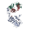 3be1S S: Starting model for refinement |
|---|---|
| Similar structure data |

-Links
 PDBj
PDBj


- Assembly
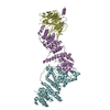
| Deposited unit | 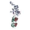
| ||||||||
|---|---|---|---|---|---|---|---|---|---|
| 1 |
| ||||||||
| Unit cell |
|
-Components
| #1: Protein | Mass: 69420.812 Da / Num. of mol.: 1 / Fragment: UNP residues 23-652 Source method: isolated from a genetically manipulated source Source: (gene. exp.) Homo sapiens (human) / Cell line: HEK293 / Gene: ERBB2, HER2, MLN19, NEU, NGL / Production host: Mammalian expression vector pBGSA (others)References: UniProt: P04626, receptor protein-tyrosine kinase | ||||
|---|---|---|---|---|---|
| #2: Antibody | Mass: 23868.588 Da / Num. of mol.: 1 Source method: isolated from a genetically manipulated source Details: Vh and Ch1 domains of antibody heavy chain / Source: (gene. exp.) Homo sapiens (human) / Cell line: HEK293 / Cell line (production host): HEK293 / Production host: Homo sapiens (human) | ||||
| #3: Antibody | Mass: 24161.936 Da / Num. of mol.: 1 Source method: isolated from a genetically manipulated source Details: Antibody, light chain / Source: (gene. exp.) Homo sapiens (human) / Cell line (production host): HEK293 / Production host: Homo sapiens (human) | ||||
| #4: Polysaccharide | Source method: isolated from a genetically manipulated source #5: Sugar | ChemComp-NAG / |   Type: D-saccharide, beta linking / Mass: 221.208 Da / Num. of mol.: 1 Type: D-saccharide, beta linking / Mass: 221.208 Da / Num. of mol.: 1Source method: isolated from a genetically manipulated source Formula: C8H15NO6 Has protein modification | Y | |
-Experimental details
-Experiment
| Experiment | Method: X-RAY DIFFRACTION / Number of used crystals: 1 |
|---|
- Sample preparation
| Crystal | Density Matthews: 5.4 Å3/Da / Density % sol: 77.22 % |
|---|---|
| Crystal grow | Temperature: 295 K / Method: vapor diffusion / pH: 6.5 / Details: PEG8000, ethylene glycol, MES buffer |
-Data collection
| Diffraction | Mean temperature: 100 K |
|---|---|
| Diffraction source | Source: SYNCHROTRON / Site: SLS  / Beamline: X06DA / Wavelength: 1 Å / Beamline: X06DA / Wavelength: 1 Å |
| Detector | Type: DECTRIS PILATUS3 X 1M / Detector: PIXEL / Date: Oct 25, 2015 |
| Radiation | Protocol: SINGLE WAVELENGTH / Monochromatic (M) / Laue (L): M / Scattering type: x-ray |
| Radiation wavelength | Wavelength: 1 Å / Relative weight: 1 |
| Reflection | Resolution: 3.77→40 Å / Num. obs: 25241 / % possible obs: 99.94 % / Redundancy: 12.4 % / Biso Wilson estimate: 80 Å2 / Rsym value: 0.13 / Net I/σ(I): 12 |
| Reflection shell | Resolution: 3.77→3.87 Å / Redundancy: 10.6 % / Rsym value: 0.87 / % possible all: 95 |
- Processing
| Software |
| ||||||||||||||||||||||||||||||||||||||||||||||||||||||||||||||||||||||||||||||||||||||||||||||||||||||||||||||||||||||||||||||||||||||||||||||||||||||||||||||||||||||||||||||||||||||
|---|---|---|---|---|---|---|---|---|---|---|---|---|---|---|---|---|---|---|---|---|---|---|---|---|---|---|---|---|---|---|---|---|---|---|---|---|---|---|---|---|---|---|---|---|---|---|---|---|---|---|---|---|---|---|---|---|---|---|---|---|---|---|---|---|---|---|---|---|---|---|---|---|---|---|---|---|---|---|---|---|---|---|---|---|---|---|---|---|---|---|---|---|---|---|---|---|---|---|---|---|---|---|---|---|---|---|---|---|---|---|---|---|---|---|---|---|---|---|---|---|---|---|---|---|---|---|---|---|---|---|---|---|---|---|---|---|---|---|---|---|---|---|---|---|---|---|---|---|---|---|---|---|---|---|---|---|---|---|---|---|---|---|---|---|---|---|---|---|---|---|---|---|---|---|---|---|---|---|---|---|---|---|---|
| Refinement | Method to determine structure: MOLECULAR REPLACEMENT Starting model: 3BE1 Resolution: 3.77→40 Å / Cor.coef. Fo:Fc: 0.902 / Cor.coef. Fo:Fc free: 0.883 / SU B: 36.28 / SU ML: 0.507 / Cross valid method: THROUGHOUT / ESU R: 2.035 / ESU R Free: 0.617 / Details: HYDROGENS HAVE BEEN ADDED IN THE RIDING POSITIONS
| ||||||||||||||||||||||||||||||||||||||||||||||||||||||||||||||||||||||||||||||||||||||||||||||||||||||||||||||||||||||||||||||||||||||||||||||||||||||||||||||||||||||||||||||||||||||
| Solvent computation | Ion probe radii: 0.8 Å / Shrinkage radii: 0.8 Å / VDW probe radii: 1.2 Å | ||||||||||||||||||||||||||||||||||||||||||||||||||||||||||||||||||||||||||||||||||||||||||||||||||||||||||||||||||||||||||||||||||||||||||||||||||||||||||||||||||||||||||||||||||||||
| Displacement parameters | Biso mean: 77.221 Å2
| ||||||||||||||||||||||||||||||||||||||||||||||||||||||||||||||||||||||||||||||||||||||||||||||||||||||||||||||||||||||||||||||||||||||||||||||||||||||||||||||||||||||||||||||||||||||
| Refinement step | Cycle: 1 / Resolution: 3.77→40 Å
| ||||||||||||||||||||||||||||||||||||||||||||||||||||||||||||||||||||||||||||||||||||||||||||||||||||||||||||||||||||||||||||||||||||||||||||||||||||||||||||||||||||||||||||||||||||||
| Refine LS restraints |
|