 Movie
Movie Controller


+ Open data

- Basic information
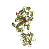
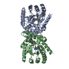
| Entry | Database: PDB / ID: 2ffv | ||||||
|---|---|---|---|---|---|---|---|
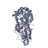
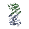
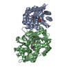
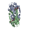
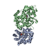
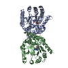
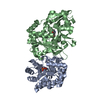
| Title | Human ppGalNAcT-2 complexed with manganese and UDP | ||||||
 Components Components | Polypeptide N-acetylgalactosaminyltransferase 2 | ||||||
 Keywords Keywords | TRANSFERASE / ppGalNAcT / mucin / glycosyltransferase | ||||||
| Function / homology |  Function and homology information Function and homology informationpolypeptide N-acetylgalactosaminyltransferase / polypeptide N-acetylgalactosaminyltransferase activity / protein O-linked glycosylation via N-acetylgalactosamine / O-linked glycosylation of mucins / protein O-linked glycosylation / Golgi stack / COPI-independent Golgi-to-ER retrograde traffic / Golgi cisterna membrane / positive regulation of immunoglobulin production / protein maturation ...polypeptide N-acetylgalactosaminyltransferase / polypeptide N-acetylgalactosaminyltransferase activity / protein O-linked glycosylation via N-acetylgalactosamine / O-linked glycosylation of mucins / protein O-linked glycosylation / Golgi stack / COPI-independent Golgi-to-ER retrograde traffic / Golgi cisterna membrane / positive regulation of immunoglobulin production / protein maturation / manganese ion binding / carbohydrate binding / Golgi membrane / endoplasmic reticulum membrane / perinuclear region of cytoplasm / Golgi apparatus / extracellular region / membrane Similarity search - Function | ||||||
| Biological species |  Homo sapiens (human) Homo sapiens (human) | ||||||
| Method |  X-RAY DIFFRACTION / SYNCHROTRON / MOLECULAR REPLACEMENT / Resolution: 2.75 Å X-RAY DIFFRACTION / SYNCHROTRON / MOLECULAR REPLACEMENT / Resolution: 2.75 Å | ||||||
 Authors Authors | Fritz, T.A. | ||||||
 Citation Citation | Journal: J.Biol.Chem. / Year: 2006 Title: Dynamic Association between the Catalytic and Lectin Domains of Human UDP-GalNAc:Polypeptide {alpha}-N-Acetylgalactosaminyltransferase-2 Authors: Fritz, T.A. / Raman, J. / Tabak, L.A. | ||||||
| History |
|
- Structure visualization
| Structure viewer | Molecule: MolmilJmol/JSmol |
|---|
- Downloads & links
-Download
| PDBx/mmCIF format | 2ffv.cif.gz | 206.4 KB | Display | PDBx/mmCIF format |
|---|---|---|---|---|
| PDB format | pdb2ffv.ent.gz | 162.8 KB | Display | PDB format |
| PDBx/mmJSON format | 2ffv.json.gz | Tree view | PDBx/mmJSON format | |
| Others |  Other downloads Other downloads |
-Validation report
| Arichive directory | https://data.pdbj.org/pub/pdb/validation_reports/ff/2ffvftp://data.pdbj.org/pub/pdb/validation_reports/ff/2ffv | HTTPS FTP |
|---|
-Related structure data
| Related structure data | 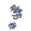 2ffuC 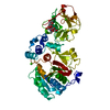 1xhbS S: Starting model for refinement C: citing same article ( |
|---|---|
| Similar structure data |

-Links
 PDBj
PDBj


- Assembly
| Deposited unit | 
| ||||||||
|---|---|---|---|---|---|---|---|---|---|
| 1 | 
| ||||||||
| 2 | 
| ||||||||
| 3 | 
| ||||||||
| Unit cell |
| ||||||||
| Details | The biological assembly is either one of the two monomers |
-Components
| #1: Protein | Mass: 57129.844 Da / Num. of mol.: 2 / Fragment: Catalytic and lectin domains Source method: isolated from a genetically manipulated source Source: (gene. exp.) Homo sapiens (human)Description: Invitrogen pPIC9 vector with TEV-protease-cleavable, N-terminal engineered 6His tag Gene: GALNT2 / Plasmid: pPIC9 / Production host:  Pichia pastoris (fungus) / Strain (production host): SMD1168 Pichia pastoris (fungus) / Strain (production host): SMD1168References: UniProt: Q10471, polypeptide N-acetylgalactosaminyltransferase #2: Chemical |   Mass: 54.938 Da / Num. of mol.: 2 / Source method: obtained synthetically / Formula: Mn Mass: 54.938 Da / Num. of mol.: 2 / Source method: obtained synthetically / Formula: Mn#3: Chemical |   Type: RNA linking / Mass: 404.161 Da / Num. of mol.: 2 / Source method: obtained synthetically / Formula: C9H14N2O12P2 / Comment: UDP*YM Type: RNA linking / Mass: 404.161 Da / Num. of mol.: 2 / Source method: obtained synthetically / Formula: C9H14N2O12P2 / Comment: UDP*YMHas protein modification | Y | |
|---|
-Experimental details
-Experiment
| Experiment | Method: X-RAY DIFFRACTION / Number of used crystals: 1 |
|---|
- Sample preparation
| Crystal | Density Matthews: 2.83 Å3/Da / Density % sol: 56.51 % |
|---|---|
| Crystal grow | Temperature: 298 K / Method: vapor diffusion, hanging drop / pH: 7 Details: PEG, Hepes, MnCl2, EDTA, mercaptoethanol, UDP, pH 7.0, VAPOR DIFFUSION, HANGING DROP, temperature 298K |
-Data collection
| Diffraction | Mean temperature: 95 K |
|---|---|
| Diffraction source | Source: SYNCHROTRON / Site: APS  / Beamline: 22-ID / Wavelength: 0.97472 Å / Beamline: 22-ID / Wavelength: 0.97472 Å |
| Detector | Type: MARMOSAIC 300 mm CCD / Detector: CCD / Date: Jul 2, 2005 |
| Radiation | Protocol: SINGLE WAVELENGTH / Monochromatic (M) / Laue (L): M / Scattering type: x-ray |
| Radiation wavelength | Wavelength: 0.97472 Å / Relative weight: 1 |
| Reflection | Resolution: 2.75→49.1 Å / Num. all: 34646 / Num. obs: 34265 / % possible obs: 98.9 % / Observed criterion σ(I): -3 / Biso Wilson estimate: 64.6 Å2 |
- Processing
| Software |
| ||||||||||||||||||||||||||||||||||||
|---|---|---|---|---|---|---|---|---|---|---|---|---|---|---|---|---|---|---|---|---|---|---|---|---|---|---|---|---|---|---|---|---|---|---|---|---|---|
| Refinement | Method to determine structure: MOLECULAR REPLACEMENT Starting model: Separate catalytic domains of PDB entry 1XHB in which nonconserved residues were changed to alanine Resolution: 2.75→49.1 Å / Rfactor Rfree error: 0.007 / Data cutoff high absF: 2290873.5 / Data cutoff low absF: 0 / Isotropic thermal model: RESTRAINED / Cross valid method: THROUGHOUT / σ(F): 0 / Stereochemistry target values: Engh & Huber
| ||||||||||||||||||||||||||||||||||||
| Solvent computation | Solvent model: FLAT MODEL / Bsol: 39.276 Å2 / ksol: 0.334354 e/Å3 | ||||||||||||||||||||||||||||||||||||
| Displacement parameters | Biso mean: 44.6 Å2
| ||||||||||||||||||||||||||||||||||||
| Refine analyze |
| ||||||||||||||||||||||||||||||||||||
| Refinement step | Cycle: LAST / Resolution: 2.75→49.1 Å
| ||||||||||||||||||||||||||||||||||||
| Refine LS restraints |
| ||||||||||||||||||||||||||||||||||||
| LS refinement shell | Resolution: 2.75→2.92 Å / Rfactor Rfree error: 0.026 / Total num. of bins used: 6
| ||||||||||||||||||||||||||||||||||||
| Xplor file |
|
