 Movie
Movie Controller

[English] 日本語
 Yorodumi
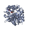
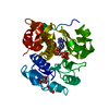
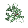
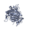
- PDB-2ffi: Crystal Structure of Putative 2-Pyrone-4,6-Dicarboxylic Acid Hydr... -
+ Open data

- Basic information
| Entry | Database: PDB / ID: 2ffi | ||||||
|---|---|---|---|---|---|---|---|
| Title | Crystal Structure of Putative 2-Pyrone-4,6-Dicarboxylic Acid Hydrolase from Pseudomonas putida, Northeast Structural Genomics Target PpR23. | ||||||
 Components Components | 2-pyrone-4,6-dicarboxylic acid hydrolase, putative | ||||||
 Keywords Keywords | STRUCTURAL GENOMICS / UNKNOWN FUNCTION / TIM-barrel protein. / PSI / Protein Structure Initiative / Northeast Structural Genomics Consortium / NESG | ||||||
| Function / homology |  Function and homology information Function and homology information2-pyrone-4,6-dicarboxylate lactonase / 2-pyrone-4,6-dicarboxylate lactonase activity Similarity search - Function | ||||||
| Biological species |  Pseudomonas putida (bacteria) Pseudomonas putida (bacteria) | ||||||
| Method |  X-RAY DIFFRACTION / SYNCHROTRON / MAD / Resolution: 2.61 Å X-RAY DIFFRACTION / SYNCHROTRON / MAD / Resolution: 2.61 Å | ||||||
 Authors Authors | Forouhar, F. / Su, M. / Jayaraman, S. / Conover, K. / Xiao, R. / Acton, T.B. / Montelione, G.T. / Hunt, J.F. / Tong, L. / Northeast Structural Genomics Consortium (NESG) | ||||||
 Citation Citation | Journal: To be Published Title: Crystal Structure of Putative 2-Pyrone-4,6-Dicarboxylic Acid Hydrolase from Pseudomonas putida, Northeast Structural Genomics Target PpR23. Authors: Forouhar, F. / Su, M. / Jayaraman, S. / Conover, K. / Xiao, R. / Acton, T.B. / Montelione, G.T. / Hunt, J.F. / Tong, L. / Northeast Structural Genomics Consortium (NESG) | ||||||
| History |
|
- Structure visualization
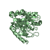
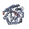
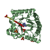
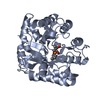
| Structure viewer | Molecule: MolmilJmol/JSmol |
|---|
- Downloads & links
-Download
| PDBx/mmCIF format | 2ffi.cif.gz | 117.6 KB | Display | PDBx/mmCIF format |
|---|---|---|---|---|
| PDB format | pdb2ffi.ent.gz | 91.4 KB | Display | PDB format |
| PDBx/mmJSON format | 2ffi.json.gz | Tree view | PDBx/mmJSON format | |
| Others |  Other downloads Other downloads |
-Validation report
| Summary document | 2ffi_validation.pdf.gz | 450.6 KB | Display | wwPDB validaton report |
|---|---|---|---|---|
| Full document | 2ffi_full_validation.pdf.gz | 473.4 KB | Display | |
| Data in XML | 2ffi_validation.xml.gz | 26.4 KB | Display | |
| Data in CIF | 2ffi_validation.cif.gz | 34.2 KB | Display | |
| Arichive directory | https://data.pdbj.org/pub/pdb/validation_reports/ff/2ffiftp://data.pdbj.org/pub/pdb/validation_reports/ff/2ffi | HTTPS FTP |
-Related structure data
| Similar structure data | |
|---|---|
| Other databases |


-Links
 PDBj
PDBj

- Assembly
| Deposited unit | 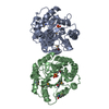
| ||||||||
|---|---|---|---|---|---|---|---|---|---|
| 1 | 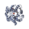
| ||||||||
| 2 | 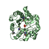
| ||||||||
| Unit cell |
|
-Components
| #1: Protein | Mass: 32427.701 Da / Num. of mol.: 2 Source method: isolated from a genetically manipulated source Source: (gene. exp.) Pseudomonas putida (bacteria) / Strain: KT2440 / Gene: PP1699 / Plasmid: pET2 / Production host: #2: Chemical |   Mass: 94.971 Da / Num. of mol.: 2 / Source method: obtained synthetically / Formula: PO4 Mass: 94.971 Da / Num. of mol.: 2 / Source method: obtained synthetically / Formula: PO4#3: Water | ChemComp-HOH / |  Mass: 18.015 Da / Num. of mol.: 55 / Source method: isolated from a natural source / Formula: H2O Mass: 18.015 Da / Num. of mol.: 55 / Source method: isolated from a natural source / Formula: H2OHas protein modification | Y | |
|---|
-Experimental details
-Experiment
| Experiment | Method: X-RAY DIFFRACTION / Number of used crystals: 1 |
|---|
- Sample preparation
| Crystal | Density Matthews: 1.89 Å3/Da / Density % sol: 34.94 % |
|---|---|
| Crystal grow | Temperature: 291 K / Method: vapor diffusion, hanging drop / pH: 4.9 Details: 100mM Na Acetate, 22.1% PEG3350, 100 mM KH2PO4, and 5 mM DTT, pH 4.9, VAPOR DIFFUSION, HANGING DROP, temperature 291K |
-Data collection
| Diffraction | Mean temperature: 100 K | ||||||||||||
|---|---|---|---|---|---|---|---|---|---|---|---|---|---|
| Diffraction source | Source: SYNCHROTRON / Site: NSLS  / Beamline: X4A / Wavelength: 0.9790, 0.9795, 0.9670 / Beamline: X4A / Wavelength: 0.9790, 0.9795, 0.9670 | ||||||||||||
| Detector | Type: ADSC QUANTUM 4 / Detector: CCD / Date: Oct 4, 2005 / Details: Mirrors. | ||||||||||||
| Radiation | Monochromator: Si 111 CHANNEL / Protocol: MAD / Monochromatic (M) / Laue (L): M / Scattering type: x-ray | ||||||||||||
| Radiation wavelength |
| ||||||||||||
| Reflection | Resolution: 2.6→28.91 Å / Num. all: 28897 / Num. obs: 28492 / % possible obs: 98.6 % / Observed criterion σ(F): 0 / Observed criterion σ(I): 0 / Redundancy: 3.5 % / Biso Wilson estimate: 18.5 Å2 / Rmerge(I) obs: 0.133 / Rsym value: 0.112 / Net I/σ(I): 8.95 | ||||||||||||
| Reflection shell | Resolution: 2.6→2.69 Å / Redundancy: 3.4 % / Rmerge(I) obs: 0.451 / Mean I/σ(I) obs: 3 / Num. unique all: 1403 / Rsym value: 0.364 / % possible all: 97.5 |
- Processing
| Software |
| |||||||||||||||||||||||||
|---|---|---|---|---|---|---|---|---|---|---|---|---|---|---|---|---|---|---|---|---|---|---|---|---|---|---|
| Refinement | Method to determine structure: MAD / Resolution: 2.61→28.91 Å / Rfactor Rfree error: 0.006 / Data cutoff high absF: 259422.81 / Data cutoff low absF: 0 / Isotropic thermal model: OVERALL / Cross valid method: THROUGHOUT / σ(F): 2 / σ(I): 2 / Stereochemistry target values: Engh & Huber
| |||||||||||||||||||||||||
| Solvent computation | Solvent model: FLAT MODEL / Bsol: 16.5855 Å2 / ksol: 0.294793 e/Å3 | |||||||||||||||||||||||||
| Displacement parameters | Biso mean: 25.9 Å2
| |||||||||||||||||||||||||
| Refine analyze |
| |||||||||||||||||||||||||
| Refinement step | Cycle: LAST / Resolution: 2.61→28.91 Å
| |||||||||||||||||||||||||
| Refine LS restraints |
| |||||||||||||||||||||||||
| LS refinement shell | Resolution: 2.6→2.76 Å / Rfactor Rfree error: 0.022 / Total num. of bins used: 6
|
