| 登録情報 | データベース: PDB / ID: 2ff6
|
|---|
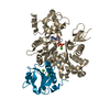
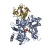
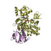
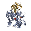
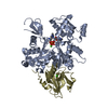
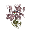
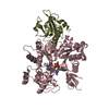
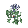
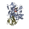
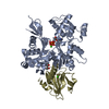
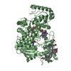
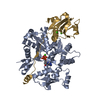
| タイトル | Crystal structure of Gelsolin domain 1:ciboulot domain 2 hybrid in complex with actin |
|---|
 要素 要素 | - Actin, alpha skeletal muscle
- CG4944-PC, isoform C
- Gelsolin
|
|---|
 キーワード キーワード | STRUCTURAL PROTEIN/CONTRACTILE PROTEIN / Protein-Protein complex / STRUCTURAL PROTEIN-CONTRACTILE PROTEIN COMPLEX |
|---|
| 機能・相同性 |  機能・相同性情報 機能・相同性情報
larval central nervous system remodeling / striated muscle atrophy / regulation of establishment of T cell polarity / regulation of plasma membrane raft polarization / regulation of receptor clustering / positive regulation of keratinocyte apoptotic process / renal protein absorption / positive regulation of protein processing in phagocytic vesicle / phosphatidylinositol 3-kinase catalytic subunit binding / positive regulation of actin nucleation ...larval central nervous system remodeling / striated muscle atrophy / regulation of establishment of T cell polarity / regulation of plasma membrane raft polarization / regulation of receptor clustering / positive regulation of keratinocyte apoptotic process / renal protein absorption / positive regulation of protein processing in phagocytic vesicle / phosphatidylinositol 3-kinase catalytic subunit binding / positive regulation of actin nucleation / actin cap / myosin II binding / host-mediated suppression of symbiont invasion / actin filament severing / barbed-end actin filament capping / cell projection assembly / actin polymerization or depolymerization / actin filament depolymerization / actin filament capping / relaxation of cardiac muscle / Sensory processing of sound by outer hair cells of the cochlea / phagocytosis, engulfment / cardiac muscle cell contraction / cytoskeletal motor activator activity / myosin heavy chain binding / hepatocyte apoptotic process / tropomyosin binding / actin filament bundle / troponin I binding / filamentous actin / mesenchyme migration / skeletal muscle myofibril / actin filament bundle assembly / sarcoplasm / striated muscle thin filament / skeletal muscle thin filament assembly / cilium assembly / actin monomer binding / Caspase-mediated cleavage of cytoskeletal proteins / phagocytic vesicle / skeletal muscle fiber development / response to muscle stretch / stress fiber / titin binding / phosphatidylinositol-4,5-bisphosphate binding / actin filament polymerization / central nervous system development / actin filament organization / filopodium / actin filament / brain development / cellular response to type II interferon / protein destabilization / 加水分解酵素; 酸無水物に作用; 酸無水物に作用・細胞または細胞小器官の運動に関与 / calcium-dependent protein binding / actin filament binding / lamellipodium / actin cytoskeleton / actin binding / cell body / secretory granule lumen / blood microparticle / amyloid fibril formation / ficolin-1-rich granule lumen / cytoskeleton / Amyloid fiber formation / protein domain specific binding / focal adhesion / hydrolase activity / calcium ion binding / Neutrophil degranulation / positive regulation of gene expression / magnesium ion binding / : / extracellular exosome / extracellular region / ATP binding / identical protein binding / plasma membrane / cytoplasm / cytosol類似検索 - 分子機能 Beta-thymosin / Beta-thymosin superfamily / Thymosin beta-4 family / Thymosin beta actin-binding motif. / Villin/Gelsolin / Gelsolin homology domain / Severin / Severin / Gelsolin-like domain / Gelsolin repeat ...Beta-thymosin / Beta-thymosin superfamily / Thymosin beta-4 family / Thymosin beta actin-binding motif. / Villin/Gelsolin / Gelsolin homology domain / Severin / Severin / Gelsolin-like domain / Gelsolin repeat / ADF-H/Gelsolin-like domain superfamily / ATPase, substrate binding domain, subdomain 4 / Actin; Chain A, domain 4 / ATPase, nucleotide binding domain / Actins signature 1. / Actin, conserved site / Actins signature 2. / Actin/actin-like conserved site / Actins and actin-related proteins signature. / Actin / Actin family / Actin / ATPase, nucleotide binding domain / Nucleotidyltransferase; domain 5 / Alpha-Beta Complex / 2-Layer Sandwich / 3-Layer(aba) Sandwich / Alpha Beta類似検索 - ドメイン・相同性 ADENOSINE-5'-TRIPHOSPHATE / Gelsolin / Actin, alpha skeletal muscle / Ciboulot, isoform C類似検索 - 構成要素 |
|---|
| 生物種 |  Homo sapiens (ヒト) Homo sapiens (ヒト)
  Drosophila melanogaster (キイロショウジョウバエ) Drosophila melanogaster (キイロショウジョウバエ)
  Oryctolagus cuniculus (ウサギ) Oryctolagus cuniculus (ウサギ) |
|---|
| 手法 |  X線回折 / シンクロトロン / 分子置換 / 解像度: 2.05 Å X線回折 / シンクロトロン / 分子置換 / 解像度: 2.05 Å |
|---|
 データ登録者 データ登録者 | Aguda, A.H. / Xue, B. / Robinson, R.C. |
|---|
 引用 引用 | ジャーナル: Structure / 年: 2006
タイトル: The Structural Basis of Actin Interaction with Multiple WH2/beta-Thymosin Motif-Containing Proteins
著者: Aguda, A.H. / Xue, B. / Irobi, E. / Preat, T. / Robinson, R.C. |
|---|
| 履歴 | | 登録 | 2005年12月19日 | 登録サイト: RCSB / 処理サイト: PDBJ |
|---|
| 改定 1.0 | 2006年3月21日 | Provider: repository / タイプ: Initial release |
|---|
| 改定 1.1 | 2008年5月1日 | Group: Version format compliance |
|---|
| 改定 1.2 | 2011年7月13日 | Group: Version format compliance |
|---|
| 改定 1.3 | 2018年5月23日 | Group: Data collection / カテゴリ: diffrn_source / Item: _diffrn_source.pdbx_synchrotron_site |
|---|
| 改定 1.4 | 2024年11月6日 | Group: Data collection / Database references ...Data collection / Database references / Derived calculations / Structure summary
カテゴリ: chem_comp_atom / chem_comp_bond ...chem_comp_atom / chem_comp_bond / database_2 / pdbx_entry_details / pdbx_modification_feature / pdbx_struct_conn_angle / struct_conn / struct_ref_seq_dif / struct_site
Item: _database_2.pdbx_DOI / _database_2.pdbx_database_accession ..._database_2.pdbx_DOI / _database_2.pdbx_database_accession / _pdbx_struct_conn_angle.ptnr1_auth_asym_id / _pdbx_struct_conn_angle.ptnr1_auth_comp_id / _pdbx_struct_conn_angle.ptnr1_auth_seq_id / _pdbx_struct_conn_angle.ptnr1_label_asym_id / _pdbx_struct_conn_angle.ptnr1_label_atom_id / _pdbx_struct_conn_angle.ptnr1_label_comp_id / _pdbx_struct_conn_angle.ptnr1_label_seq_id / _pdbx_struct_conn_angle.ptnr2_auth_asym_id / _pdbx_struct_conn_angle.ptnr2_auth_seq_id / _pdbx_struct_conn_angle.ptnr2_label_asym_id / _pdbx_struct_conn_angle.ptnr3_auth_asym_id / _pdbx_struct_conn_angle.ptnr3_auth_comp_id / _pdbx_struct_conn_angle.ptnr3_auth_seq_id / _pdbx_struct_conn_angle.ptnr3_label_asym_id / _pdbx_struct_conn_angle.ptnr3_label_atom_id / _pdbx_struct_conn_angle.ptnr3_label_comp_id / _pdbx_struct_conn_angle.ptnr3_label_seq_id / _pdbx_struct_conn_angle.value / _struct_conn.pdbx_dist_value / _struct_conn.pdbx_leaving_atom_flag / _struct_conn.ptnr1_auth_asym_id / _struct_conn.ptnr1_auth_comp_id / _struct_conn.ptnr1_auth_seq_id / _struct_conn.ptnr1_label_asym_id / _struct_conn.ptnr1_label_atom_id / _struct_conn.ptnr1_label_comp_id / _struct_conn.ptnr1_label_seq_id / _struct_conn.ptnr2_auth_asym_id / _struct_conn.ptnr2_auth_comp_id / _struct_conn.ptnr2_auth_seq_id / _struct_conn.ptnr2_label_asym_id / _struct_conn.ptnr2_label_atom_id / _struct_conn.ptnr2_label_comp_id / _struct_conn.ptnr2_label_seq_id / _struct_ref_seq_dif.details / _struct_site.pdbx_auth_asym_id / _struct_site.pdbx_auth_comp_id / _struct_site.pdbx_auth_seq_id |
|---|
|
|---|
 ムービー
ムービー コントローラー


 データを開く
 基本情報
基本情報 構造の表示
構造の表示 ダウンロードとリンク
 その他のダウンロード

 PDBj
PDBj


 集合体
集合体


 分子量: 40.078 Da / 分子数: 3 / 由来タイプ: 合成 / 式: Ca
 分子量: 507.181 Da / 分子数: 1 / 由来タイプ: 合成 / 式: C10H16N5O13P3 / コメント: ATP, エネルギー貯蔵分子*YM
 試料調製
試料調製 / ビームライン: I711 / 波長: 0.934 Å
 解析
解析