 Movie
Movie Controller

[English] 日本語
 Yorodumi
- PDB-2f2p: Structure of calmodulin bound to a calcineurin peptide: a new way... -
+ Open data

- Basic information
| Entry | Database: PDB / ID: 2f2p | ||||||
|---|---|---|---|---|---|---|---|
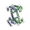
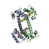
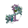
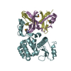
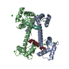
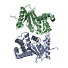
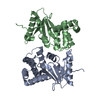
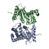
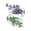
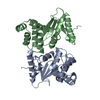
| Title | Structure of calmodulin bound to a calcineurin peptide: a new way of making an old binding mode | ||||||
 Components Components | Calmodulin fused with calmodulin-binding domain of calcineurin | ||||||
 Keywords Keywords | METAL BINDING PROTEIN / EF-hands / calcium / calmodulin / calcineurin | ||||||
| Function / homology |  Function and homology information Function and homology informationCLEC7A (Dectin-1) induces NFAT activation / Calcineurin activates NFAT / FCERI mediated Ca+2 mobilization / regulation of cell proliferation involved in kidney morphogenesis / positive regulation of glomerulus development / negative regulation of signaling / positive regulation of saliva secretion / calmodulin-dependent protein phosphatase activity / calcineurin complex / renal filtration ...CLEC7A (Dectin-1) induces NFAT activation / Calcineurin activates NFAT / FCERI mediated Ca+2 mobilization / regulation of cell proliferation involved in kidney morphogenesis / positive regulation of glomerulus development / negative regulation of signaling / positive regulation of saliva secretion / calmodulin-dependent protein phosphatase activity / calcineurin complex / renal filtration / calcineurin-NFAT signaling cascade / positive regulation of osteoclast differentiation / dephosphorylation / Ca2+ pathway / protein-serine/threonine phosphatase / positive regulation of activated T cell proliferation / epidermis development / positive regulation of osteoblast differentiation / protein dephosphorylation / keratinocyte differentiation / skeletal muscle fiber development / regulation of release of sequestered calcium ion into cytosol by sarcoplasmic reticulum / response to calcium ion / sarcolemma / modulation of chemical synaptic transmission / Z disc / spindle pole / myelin sheath / dendritic spine / calmodulin binding / protein domain specific binding / calcium ion binding / centrosome / protein-containing complex / metal ion binding / cytoplasm / cytosol Similarity search - Function | ||||||
| Biological species |  | ||||||
| Method |  X-RAY DIFFRACTION / SYNCHROTRON / MOLECULAR REPLACEMENT / Resolution: 2.6 Å X-RAY DIFFRACTION / SYNCHROTRON / MOLECULAR REPLACEMENT / Resolution: 2.6 Å | ||||||
 Authors Authors | Ye, Q. / Wong, A. / Jia, Z. | ||||||
 Citation Citation | Journal: Biochemistry / Year: 2006 Title: Structure of calmodulin bound to a calcineurin Peptide: a new way of making an old binding mode. Authors: Ye, Q. / Li, X. / Wong, A. / Wei, Q. / Jia, Z. | ||||||
| History |
|
- Structure visualization
| Structure viewer | Molecule: MolmilJmol/JSmol |
|---|
- Downloads & links
-Download
| PDBx/mmCIF format | 2f2p.cif.gz | 81.8 KB | Display | PDBx/mmCIF format |
|---|---|---|---|---|
| PDB format | pdb2f2p.ent.gz | 61.3 KB | Display | PDB format |
| PDBx/mmJSON format | 2f2p.json.gz | Tree view | PDBx/mmJSON format | |
| Others |  Other downloads Other downloads |
-Validation report
| Arichive directory | https://data.pdbj.org/pub/pdb/validation_reports/f2/2f2pftp://data.pdbj.org/pub/pdb/validation_reports/f2/2f2p | HTTPS FTP |
|---|
-Related structure data
| Related structure data |  2f2oC 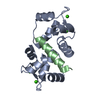 1cdmS C: citing same article ( S: Starting model for refinement |
|---|---|
| Similar structure data |
-Links
 PDBj
PDBj

- Assembly
| Deposited unit | 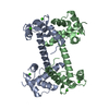
| ||||||||
|---|---|---|---|---|---|---|---|---|---|
| 1 |
| ||||||||
| 2 | 
| ||||||||
| Unit cell |
|
-Components
| #1: Protein | Mass: 19925.254 Da / Num. of mol.: 2 Source method: isolated from a genetically manipulated source Source: (gene. exp.)  #2: Chemical | ChemComp-CA /   Mass: 40.078 Da / Num. of mol.: 8 / Source method: obtained synthetically / Formula: Ca Mass: 40.078 Da / Num. of mol.: 8 / Source method: obtained synthetically / Formula: Ca#3: Water | ChemComp-HOH / |  Mass: 18.015 Da / Num. of mol.: 29 / Source method: isolated from a natural source / Formula: H2O Mass: 18.015 Da / Num. of mol.: 29 / Source method: isolated from a natural source / Formula: H2O |
|---|
-Experimental details
-Experiment
| Experiment | Method: X-RAY DIFFRACTION / Number of used crystals: 1 |
|---|
- Sample preparation
| Crystal | Density Matthews: 3.22 Å3/Da / Density % sol: 61.75 % |
|---|---|
| Crystal grow | Temperature: 298 K / Method: vapor diffusion, hanging drop / Details: VAPOR DIFFUSION, HANGING DROP, temperature 298K |
-Data collection
| Diffraction | Mean temperature: 100 K |
|---|---|
| Diffraction source | Source: SYNCHROTRON / Site: NSLS  / Beamline: X8C / Beamline: X8C |
| Radiation | Protocol: SINGLE WAVELENGTH / Monochromatic (M) / Laue (L): M / Scattering type: x-ray |
| Radiation wavelength | Relative weight: 1 |
| Reflection | Resolution: 2.6→119.523 Å / Num. obs: 15901 / % possible obs: 99.5 % / Observed criterion σ(F): 0 / Observed criterion σ(I): 0 / Rsym value: 0.096 |
- Processing
| Software |
| |||||||||||||||||||||||||
|---|---|---|---|---|---|---|---|---|---|---|---|---|---|---|---|---|---|---|---|---|---|---|---|---|---|---|
| Refinement | Method to determine structure: MOLECULAR REPLACEMENT Starting model: pdb entry 1CDM Resolution: 2.6→50 Å / σ(F): 0 / Stereochemistry target values: Engh & Huber
| |||||||||||||||||||||||||
| Refinement step | Cycle: LAST / Resolution: 2.6→50 Å
|
