 Movie
Movie Controller

[English] 日本語
 Yorodumi
- PDB-2f2f: Crystal structure of cytolethal distending toxin (CDT) from Actin... -
+ Open data

- Basic information
| Entry | Database: PDB / ID: 2f2f | ||||||
|---|---|---|---|---|---|---|---|
| Title | Crystal structure of cytolethal distending toxin (CDT) from Actinobacillus actinomycetemcomitans | ||||||
 Components Components |
| ||||||
 Keywords Keywords | TOXIN / Cytolethal distending toxin / CDT / Actinobacillus actinomycetemcomitans / oligomerization / stability and toxic activity | ||||||
| Function / homology |  Function and homology information Function and homology informationcatalytic activity / cell outer membrane / toxin activity / carbohydrate binding Similarity search - Function | ||||||
| Biological species |  Aggregatibacter actinomycetemcomitans (bacteria) Aggregatibacter actinomycetemcomitans (bacteria) | ||||||
| Method |  X-RAY DIFFRACTION / SYNCHROTRON / MOLECULAR REPLACEMENT / Resolution: 2.4 Å X-RAY DIFFRACTION / SYNCHROTRON / MOLECULAR REPLACEMENT / Resolution: 2.4 Å | ||||||
 Authors Authors | Yamada, T. / Komoto, J. / Saiki, K. / Konishi, K. / Takusagawa, F. | ||||||
 Citation Citation | Journal: Protein Sci. / Year: 2006 Title: Variation of loop sequence alters stability of cytolethal distending toxin (CDT): crystal structure of CDT from Actinobacillus actinomycetemcomitans Authors: Yamada, T. / Komoto, J. / Saiki, K. / Konishi, K. / Takusagawa, F. | ||||||
| History |
| ||||||
| Remark 999 | SEQUENCE AUTHOR STATES THAT SEQUENCE IN THE PDB FILE IS CORRECT ON THE BASIS OF ELECTRON DENSITY ...SEQUENCE AUTHOR STATES THAT SEQUENCE IN THE PDB FILE IS CORRECT ON THE BASIS OF ELECTRON DENSITY MAPS AND THE OTHER CDT SEQUENCES. |
- Structure visualization
| Structure viewer | Molecule: MolmilJmol/JSmol |
|---|
- Downloads & links
-Download
| PDBx/mmCIF format | 2f2f.cif.gz | 232.6 KB | Display | PDBx/mmCIF format |
|---|---|---|---|---|
| PDB format | pdb2f2f.ent.gz | 185.9 KB | Display | PDB format |
| PDBx/mmJSON format | 2f2f.json.gz | Tree view | PDBx/mmJSON format | |
| Others |  Other downloads Other downloads |
-Validation report
| Arichive directory | https://data.pdbj.org/pub/pdb/validation_reports/f2/2f2fftp://data.pdbj.org/pub/pdb/validation_reports/f2/2f2f | HTTPS FTP |
|---|
-Related structure data
| Related structure data |  1sr4S S: Starting model for refinement |
|---|---|
| Similar structure data |


-Links
 PDBj
PDBj


- Assembly
| Deposited unit | 
| ||||||||
|---|---|---|---|---|---|---|---|---|---|
| 1 | 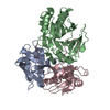
| ||||||||
| 2 | 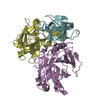
| ||||||||
| Unit cell |
| ||||||||
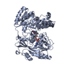
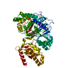
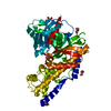
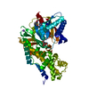
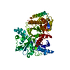
| Details | CDT is a heterotrimer, i.e., Subunit A, B, and C form a holotoxin, and Subunit D, E, and F form another holotoxin. |
-Components
| #1: Protein | Mass: 24532.748 Da / Num. of mol.: 2 Source method: isolated from a genetically manipulated source Source: (gene. exp.) Aggregatibacter actinomycetemcomitans (bacteria)Gene: cdtA / Plasmid: A19-47CDT / Production host: #2: Protein | Mass: 31528.475 Da / Num. of mol.: 2 Source method: isolated from a genetically manipulated source Source: (gene. exp.) Aggregatibacter actinomycetemcomitans (bacteria)Gene: cdtB / Plasmid: A19-47CDT / Production host: #3: Protein | Mass: 20731.672 Da / Num. of mol.: 2 Source method: isolated from a genetically manipulated source Source: (gene. exp.) Aggregatibacter actinomycetemcomitans (bacteria)Gene: cdtC / Plasmid: A19-47CDT / Production host: #4: Water | ChemComp-HOH / |  Mass: 18.015 Da / Num. of mol.: 272 / Source method: isolated from a natural source / Formula: H2O Mass: 18.015 Da / Num. of mol.: 272 / Source method: isolated from a natural source / Formula: H2OHas protein modification | Y | |
|---|
-Experimental details
-Experiment
| Experiment | Method: X-RAY DIFFRACTION / Number of used crystals: 1 |
|---|
- Sample preparation
| Crystal | Density Matthews: 2.73 Å3/Da / Density % sol: 54.89 % |
|---|---|
| Crystal grow | Temperature: 295 K / Method: vapor diffusion, hanging drop / pH: 6.5 Details: 50 mM MES, 10% MPD, 4% PEG-8000, pH 6.5, VAPOR DIFFUSION, HANGING DROP, temperature 295K |
-Data collection
| Diffraction | Mean temperature: 95 K |
|---|---|
| Diffraction source | Source: SYNCHROTRON / Site: APS  / Beamline: 19-BM / Wavelength: 1.08 Å / Beamline: 19-BM / Wavelength: 1.08 Å |
| Detector | Type: CUSTOM-MADE / Detector: CCD / Date: Jan 10, 2005 |
| Radiation | Protocol: SINGLE WAVELENGTH / Monochromatic (M) / Laue (L): M / Scattering type: x-ray |
| Radiation wavelength | Wavelength: 1.08 Å / Relative weight: 1 |
| Reflection | Resolution: 2.4→20 Å / Num. all: 66570 / Num. obs: 66570 / % possible obs: 97.2 % / Observed criterion σ(F): 0 / Observed criterion σ(I): 0 |
| Reflection shell | Resolution: 2.4→2.5 Å / % possible all: 88 |
- Processing
| Software |
| ||||||||||||||||||||
|---|---|---|---|---|---|---|---|---|---|---|---|---|---|---|---|---|---|---|---|---|---|
| Refinement | Method to determine structure: MOLECULAR REPLACEMENT Starting model: pdb entry 1SR4 Resolution: 2.4→20 Å / σ(F): 0 / Stereochemistry target values: Engh & Huber
| ||||||||||||||||||||
| Refinement step | Cycle: LAST / Resolution: 2.4→20 Å
|
