 Movie
Movie Controller

[English] 日本語
 Yorodumi
- PDB-1sr4: Crystal Structure of the Haemophilus ducreyi cytolethal distendin... -
+ Open data

- Basic information
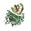
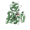
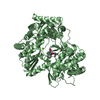
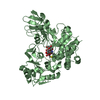
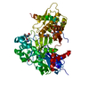
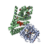
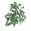
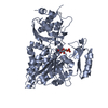
| Entry | Database: PDB / ID: 1sr4 | ||||||
|---|---|---|---|---|---|---|---|
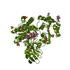
| Title | Crystal Structure of the Haemophilus ducreyi cytolethal distending toxin | ||||||
 Components Components |
| ||||||
 Keywords Keywords | TOXIN / bacterial / Haemophilus ducreyi / virulence / DNA Damage / genotoxin / cytotoxins / Cell Cycle / Apoptosis / lectin / Deoxyribonuclease I | ||||||
| Function / homology |  Function and homology information Function and homology informationcatalytic activity / cell outer membrane / toxin activity / carbohydrate binding Similarity search - Function | ||||||
| Biological species |  Haemophilus ducreyi (bacteria) Haemophilus ducreyi (bacteria) | ||||||
| Method |  X-RAY DIFFRACTION / SYNCHROTRON / SAD / Resolution: 2 Å X-RAY DIFFRACTION / SYNCHROTRON / SAD / Resolution: 2 Å | ||||||
 Authors Authors | Nesic, D. / Hsu, Y. / Stebbins, C.E. | ||||||
 Citation Citation | Journal: Nature / Year: 2004 Title: Assembly and Function of a Bacterial Genotoxin Authors: Nesic, D. / Hsu, Y. / Stebbins, C.E. | ||||||
| History |
|
- Structure visualization
| Structure viewer | Molecule: MolmilJmol/JSmol |
|---|
- Downloads & links
-Download
| PDBx/mmCIF format | 1sr4.cif.gz | 138.6 KB | Display | PDBx/mmCIF format |
|---|---|---|---|---|
| PDB format | pdb1sr4.ent.gz | 105.7 KB | Display | PDB format |
| PDBx/mmJSON format | 1sr4.json.gz | Tree view | PDBx/mmJSON format | |
| Others |  Other downloads Other downloads |
-Validation report
| Arichive directory | https://data.pdbj.org/pub/pdb/validation_reports/sr/1sr4ftp://data.pdbj.org/pub/pdb/validation_reports/sr/1sr4 | HTTPS FTP |
|---|
-Related structure data
| Similar structure data |
|---|

-Links
 PDBj
PDBj


- Assembly
| Deposited unit | 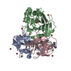
| ||||||||
|---|---|---|---|---|---|---|---|---|---|
| 1 |
| ||||||||
| Unit cell |
|
-Components
| #1: Protein | Mass: 22873.645 Da / Num. of mol.: 1 Source method: isolated from a genetically manipulated source Source: (gene. exp.) Haemophilus ducreyi (bacteria) / Gene: CDTA, HD0902 / Production host: | ||||
|---|---|---|---|---|---|
| #2: Protein | Mass: 28989.502 Da / Num. of mol.: 1 Source method: isolated from a genetically manipulated source Source: (gene. exp.) Haemophilus ducreyi (bacteria) / Production host: | ||||
| #3: Protein | Mass: 18379.771 Da / Num. of mol.: 1 Source method: isolated from a genetically manipulated source Source: (gene. exp.) Haemophilus ducreyi (bacteria) / Production host: | ||||
| #4: Chemical | ChemComp-BR /   Mass: 79.904 Da / Num. of mol.: 26 / Source method: obtained synthetically / Formula: Br Mass: 79.904 Da / Num. of mol.: 26 / Source method: obtained synthetically / Formula: Br#5: Water | ChemComp-HOH / |  Mass: 18.015 Da / Num. of mol.: 477 / Source method: isolated from a natural source / Formula: H2O Mass: 18.015 Da / Num. of mol.: 477 / Source method: isolated from a natural source / Formula: H2OHas protein modification | Y | |
-Experimental details
-Experiment
| Experiment | Method: X-RAY DIFFRACTION / Number of used crystals: 1 |
|---|
- Sample preparation
| Crystal | Density Matthews: 2.35 Å3/Da / Density % sol: 47.55 % |
|---|---|
| Crystal grow | Temperature: 296.15 K / Method: vapor diffusion, hanging drop / pH: 6.5 Details: 15-25% PEG MME 5000, 25-30% glycerol, 0.1M imidazole, 2mM DTT, pH 6.5, VAPOR DIFFUSION, HANGING DROP, temperature 296.15K |
-Data collection
| Diffraction | Mean temperature: 113.1 K |
|---|---|
| Diffraction source | Source: SYNCHROTRON / Site: NSLS  / Beamline: X9A / Wavelength: 0.92 Å / Beamline: X9A / Wavelength: 0.92 Å |
| Radiation | Monochromator: Monochromator: Double crystal monochromator with fixed exit geometry; Bragg angle range is 7.55 degrees - 28 degrees; sagitally focusing Si(111) crystals, high precision rotary energy ...Monochromator: Monochromator: Double crystal monochromator with fixed exit geometry; Bragg angle range is 7.55 degrees - 28 degrees; sagitally focusing Si(111) crystals, high precision rotary energy scale; operates in high vacuum; located 9.6 meters from the source. Mirror: For harmonics rejection and vertical focusing (0.08 mm FWHM); flat cylindrically bent mirror; independent choice of mirror angle and focal length; located 11 meters from the source Protocol: SINGLE WAVELENGTH / Monochromatic (M) / Laue (L): M / Scattering type: x-ray |
| Radiation wavelength | Wavelength: 0.92 Å / Relative weight: 1 |
| Reflection | Resolution: 2→99 Å / Num. all: 45190 / Num. obs: 45190 / % possible obs: 99.6 % / Observed criterion σ(F): 0 / Observed criterion σ(I): 0 |
| Reflection shell | Resolution: 2→2.07 Å / % possible all: 98.2 |
- Processing
| Software |
| |||||||||||||||||||||||||
|---|---|---|---|---|---|---|---|---|---|---|---|---|---|---|---|---|---|---|---|---|---|---|---|---|---|---|
| Refinement | Method to determine structure: SAD / Resolution: 2→99 Å / Cross valid method: THROUGHOUT / σ(F): 0 / Stereochemistry target values: Engh & Huber
| |||||||||||||||||||||||||
| Refinement step | Cycle: LAST / Resolution: 2→99 Å
|
