 Movie
Movie Controller


+ Open data

- Basic information
| Entry | Database: PDB / ID: 2f1t | ||||||
|---|---|---|---|---|---|---|---|
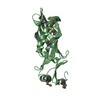
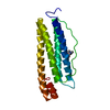
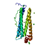
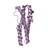
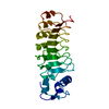
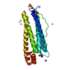
| Title | Outer membrane protein OmpW | ||||||
 Components Components | Outer membrane protein W | ||||||
 Keywords Keywords | MEMBRANE PROTEIN / outer membrane protein / beta barrel | ||||||
| Function / homology |  Function and homology information Function and homology information | ||||||
| Biological species |  | ||||||
| Method |  X-RAY DIFFRACTION / SYNCHROTRON / SIRAS / Resolution: 3 Å X-RAY DIFFRACTION / SYNCHROTRON / SIRAS / Resolution: 3 Å | ||||||
 Authors Authors | van den Berg, B. | ||||||
 Citation Citation | Journal: J.Biol.Chem. / Year: 2006 Title: The outer membrane protein OmpW forms an eight-stranded beta-barrel with a hydrophobic channel. Authors: Hong, H. / Patel, D.R. / Tamm, L.K. / van den Berg, B. | ||||||
| History |
|
- Structure visualization
| Structure viewer | Molecule: MolmilJmol/JSmol |
|---|
- Downloads & links
-Download
| PDBx/mmCIF format | 2f1t.cif.gz | 124.3 KB | Display | PDBx/mmCIF format |
|---|---|---|---|---|
| PDB format | pdb2f1t.ent.gz | 99.2 KB | Display | PDB format |
| PDBx/mmJSON format | 2f1t.json.gz | Tree view | PDBx/mmJSON format | |
| Others |  Other downloads Other downloads |
-Validation report
| Arichive directory | https://data.pdbj.org/pub/pdb/validation_reports/f1/2f1tftp://data.pdbj.org/pub/pdb/validation_reports/f1/2f1t | HTTPS FTP |
|---|
-Related structure data


-Links
 PDBj
PDBj

- Assembly
| Deposited unit | 
| ||||||||
|---|---|---|---|---|---|---|---|---|---|
| 1 | 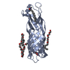
| ||||||||
| 2 | 
| ||||||||
| 3 | 
| ||||||||
| Unit cell |
|
-Components
| #1: Protein | Mass: 21700.102 Da / Num. of mol.: 3 Source method: isolated from a genetically manipulated source Source: (gene. exp.) #2: Chemical |   Mass: 229.402 Da / Num. of mol.: 3 / Source method: obtained synthetically / Formula: C14H31NO / Comment: LDAO, detergent*YM Mass: 229.402 Da / Num. of mol.: 3 / Source method: obtained synthetically / Formula: C14H31NO / Comment: LDAO, detergent*YM#3: Chemical | ChemComp-C8E / (   Mass: 306.438 Da / Num. of mol.: 8 / Source method: obtained synthetically / Formula: C16H34O5 / Comment: C8E, detergent*YM Mass: 306.438 Da / Num. of mol.: 8 / Source method: obtained synthetically / Formula: C16H34O5 / Comment: C8E, detergent*YM#4: Chemical |   Mass: 92.094 Da / Num. of mol.: 3 / Source method: obtained synthetically / Formula: C3H8O3 Mass: 92.094 Da / Num. of mol.: 3 / Source method: obtained synthetically / Formula: C3H8O3 |
|---|
-Experimental details
-Experiment
| Experiment | Method: X-RAY DIFFRACTION / Number of used crystals: 2 |
|---|
- Sample preparation
| Crystal | Density Matthews: 2.79 Å3/Da / Density % sol: 55.86 % |
|---|---|
| Crystal grow | Temperature: 295 K / Method: vapor diffusion / pH: 9 Details: 28-32% PEG400, 0.2 M CaCl2, 50 mM glycine, pH 9, VAPOR DIFFUSION, temperature 295K |
-Data collection
| Diffraction |
| ||||||||||||||||||
|---|---|---|---|---|---|---|---|---|---|---|---|---|---|---|---|---|---|---|---|
| Diffraction source |
| ||||||||||||||||||
| Detector | Type: ADSC QUANTUM 210 / Detector: CCD / Date: Jun 29, 2005 / Details: Si(111) channel cut monochromator | ||||||||||||||||||
| Radiation |
| ||||||||||||||||||
| Radiation wavelength |
| ||||||||||||||||||
| Reflection | Resolution: 2.45→46.8 Å / Num. all: 14337 / Num. obs: 14157 / % possible obs: 98.7 % / Observed criterion σ(F): 0 / Observed criterion σ(I): 2 / Redundancy: 9 % / Rmerge(I) obs: 0.067 / Net I/σ(I): 38.4 | ||||||||||||||||||
| Reflection shell | Resolution: 2.45→2.54 Å / Redundancy: 6.8 % / Rmerge(I) obs: 0.774 / Mean I/σ(I) obs: 2.1 / Num. unique all: 2678 / % possible all: 98 |

- Processing
| Software |
| |||||||||||||||||||||||||
|---|---|---|---|---|---|---|---|---|---|---|---|---|---|---|---|---|---|---|---|---|---|---|---|---|---|---|
| Refinement | Method to determine structure: SIRAS Starting model: none Resolution: 3→8 Å / Cross valid method: THROUGHOUT / σ(F): 0 / Stereochemistry target values: Engh & Huber
| |||||||||||||||||||||||||
| Displacement parameters | Biso mean: 71.39 Å2
| |||||||||||||||||||||||||
| Refine analyze |
| |||||||||||||||||||||||||
| Refinement step | Cycle: LAST / Resolution: 3→8 Å
| |||||||||||||||||||||||||
| Refine LS restraints |
| |||||||||||||||||||||||||
| LS refinement shell | Resolution: 3→3.13 Å / Rfactor Rfree error: 0.043
|
