 Movie
Movie Controller

[English] 日本語
 Yorodumi
- PDB-2ejx: Crystal structure of the hypothetical protein STK_08120 from Sulf... -
+ Open data

- Basic information
| Entry | Database: PDB / ID: 2ejx | ||||||
|---|---|---|---|---|---|---|---|
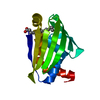
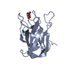
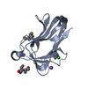
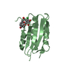
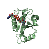
| Title | Crystal structure of the hypothetical protein STK_08120 from Sulfolobus tokodaii | ||||||
 Components Components | STK_08120 | ||||||
 Keywords Keywords | STRUCTURAL GENOMICS / UNKNOWN FUNCTION / ARCAEA / STK_08120 / NPPSFA / National Project on Protein Structural and Functional Analyses | ||||||
| Function / homology | Protein of unknown function DUF3211 / STK_08120-like / START domain / Alpha-D-Glucose-1,6-Bisphosphate; Chain A, domain 4 / START-like domain superfamily / 2-Layer Sandwich / Alpha Beta / DUF3211 domain-containing protein Function and homology information Function and homology information | ||||||
| Biological species |   Sulfolobus tokodaii str. 7 (archaea) Sulfolobus tokodaii str. 7 (archaea) | ||||||
| Method |  X-RAY DIFFRACTION / SYNCHROTRON / SAD / Resolution: 1.79 Å X-RAY DIFFRACTION / SYNCHROTRON / SAD / Resolution: 1.79 Å | ||||||
 Authors Authors | Miyakawa, T. / Miyazono, K. / Sawano, Y. / Hatano, K. / Nagata, K. / Tanokura, M. | ||||||
 Citation Citation | Journal: J.Bacteriol. / Year: 2013 Title: A thermoacidophile-specific protein family, DUF3211, functions as a fatty acid carrier with novel binding mode Authors: Miyakawa, T. / Sawano, Y. / Miyazono, K. / Miyauchi, Y. / Hatano, K. / Tanokura, M. | ||||||
| History |
|
- Structure visualization
| Structure viewer | Molecule: MolmilJmol/JSmol |
|---|
- Downloads & links
-Download
| PDBx/mmCIF format | 2ejx.cif.gz | 40.9 KB | Display | PDBx/mmCIF format |
|---|---|---|---|---|
| PDB format | pdb2ejx.ent.gz | 28.6 KB | Display | PDB format |
| PDBx/mmJSON format | 2ejx.json.gz | Tree view | PDBx/mmJSON format | |
| Others |  Other downloads Other downloads |
-Validation report
| Summary document | 2ejx_validation.pdf.gz | 427.8 KB | Display | wwPDB validaton report |
|---|---|---|---|---|
| Full document | 2ejx_full_validation.pdf.gz | 428.1 KB | Display | |
| Data in XML | 2ejx_validation.xml.gz | 8.2 KB | Display | |
| Data in CIF | 2ejx_validation.cif.gz | 10.8 KB | Display | |
| Arichive directory | https://data.pdbj.org/pub/pdb/validation_reports/ej/2ejxftp://data.pdbj.org/pub/pdb/validation_reports/ej/2ejx | HTTPS FTP |
-Related structure data


-Links
 PDBj
PDBj- Assembly
| Deposited unit | 
| ||||||||
|---|---|---|---|---|---|---|---|---|---|
| 1 |
| ||||||||
| 2 | 
| ||||||||
| Unit cell |
|
-Components
| #1: Protein | Mass: 16015.574 Da / Num. of mol.: 1 Source method: isolated from a genetically manipulated source Source: (gene. exp.) Sulfolobus tokodaii str. 7 (archaea) / Species: Sulfolobus tokodaii / Strain: strain 7 / Gene: STK_08120 / Plasmid: pET28a / Production host:  |
|---|---|
| #2: Water | ChemComp-HOH /  Mass: 18.015 Da / Num. of mol.: 102 / Source method: isolated from a natural source / Formula: H2O Mass: 18.015 Da / Num. of mol.: 102 / Source method: isolated from a natural source / Formula: H2O |
-Experimental details
-Experiment
| Experiment | Method: X-RAY DIFFRACTION / Number of used crystals: 2 |
|---|
- Sample preparation
| Crystal | Density Matthews: 2.13 Å3/Da / Density % sol: 42.14 % |
|---|---|
| Crystal grow | Temperature: 293 K / Method: vapor diffusion, sitting drop / pH: 6.6 Details: 15% PEG4000, 20% isopropanol, 100mM MES, pH 6.6, VAPOR DIFFUSION, SITTING DROP, temperature 293K |
-Data collection
| Diffraction |
| |||||||||||||||
|---|---|---|---|---|---|---|---|---|---|---|---|---|---|---|---|---|
| Diffraction source |
| |||||||||||||||
| Detector |
| |||||||||||||||
| Radiation |
| |||||||||||||||
| Radiation wavelength |
| |||||||||||||||
| Reflection | Resolution: 1.79→50 Å / Num. obs: 13524 / % possible obs: 97.8 % / Redundancy: 5.5 % / Rsym value: 0.072 / Net I/σ(I): 43.9 | |||||||||||||||
| Reflection shell | Resolution: 1.79→1.85 Å / Redundancy: 6 % / Mean I/σ(I) obs: 10.8 / Rsym value: 0.255 / % possible all: 96.6 |

- Processing
| Software |
| ||||||||||||||||||||||||||||||||||||||||||||||||||||||||||||||||||||||||||||||||||||||||||
|---|---|---|---|---|---|---|---|---|---|---|---|---|---|---|---|---|---|---|---|---|---|---|---|---|---|---|---|---|---|---|---|---|---|---|---|---|---|---|---|---|---|---|---|---|---|---|---|---|---|---|---|---|---|---|---|---|---|---|---|---|---|---|---|---|---|---|---|---|---|---|---|---|---|---|---|---|---|---|---|---|---|---|---|---|---|---|---|---|---|---|---|
| Refinement | Method to determine structure: SAD / Resolution: 1.79→20 Å / Cor.coef. Fo:Fc: 0.944 / Cor.coef. Fo:Fc free: 0.926 / SU B: 3.604 / SU ML: 0.114 / Cross valid method: THROUGHOUT / ESU R: 0.158 / ESU R Free: 0.148 / Stereochemistry target values: MAXIMUM LIKELIHOOD / Details: HYDROGENS HAVE BEEN ADDED IN THE RIDING POSITIONS
| ||||||||||||||||||||||||||||||||||||||||||||||||||||||||||||||||||||||||||||||||||||||||||
| Solvent computation | Ion probe radii: 0.8 Å / Shrinkage radii: 0.8 Å / VDW probe radii: 1.4 Å / Solvent model: MASK | ||||||||||||||||||||||||||||||||||||||||||||||||||||||||||||||||||||||||||||||||||||||||||
| Displacement parameters | Biso mean: 30.835 Å2
| ||||||||||||||||||||||||||||||||||||||||||||||||||||||||||||||||||||||||||||||||||||||||||
| Refinement step | Cycle: LAST / Resolution: 1.79→20 Å
| ||||||||||||||||||||||||||||||||||||||||||||||||||||||||||||||||||||||||||||||||||||||||||
| Refine LS restraints |
| ||||||||||||||||||||||||||||||||||||||||||||||||||||||||||||||||||||||||||||||||||||||||||
| LS refinement shell | Resolution: 1.788→1.834 Å / Total num. of bins used: 20
|
