 Movie
Movie Controller

[English] 日本語
 Yorodumi
- PDB-2eif: Eukaryotic translation initiation factor 5A from Methanococcus ja... -
+ Open data

- Basic information
| Entry | Database: PDB / ID: 2eif | ||||||
|---|---|---|---|---|---|---|---|
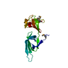
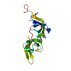
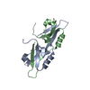
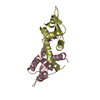
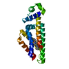
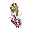
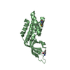
| Title | Eukaryotic translation initiation factor 5A from Methanococcus jannaschii | ||||||
 Components Components | PROTEIN (EUKARYOTIC TRANSLATION INITIATION FACTOR 5A) | ||||||
 Keywords Keywords | GENE REGULATION / EIF-5A / TRANSLATION / OB-FOLD / Structural Genomics / BSGC structure funded by NIH / Protein Structure Initiative / PSI / Berkeley Structural Genomics Center | ||||||
| Function / homology |  Function and homology information Function and homology informationtranslational elongation / translation elongation factor activity / translation initiation factor activity / ribosome binding / RNA binding / cytoplasm Similarity search - Function | ||||||
| Biological species |   Methanocaldococcus jannaschii (archaea) Methanocaldococcus jannaschii (archaea) | ||||||
| Method |  X-RAY DIFFRACTION / SYNCHROTRON / MOLECULAR REPLACEMENT / Resolution: 1.8 Å X-RAY DIFFRACTION / SYNCHROTRON / MOLECULAR REPLACEMENT / Resolution: 1.8 Å | ||||||
 Authors Authors | Kim, K.K. / Hung, L.W. / Kim, R. / Kim, S.H. / Berkeley Structural Genomics Center (BSGC) | ||||||
 Citation Citation | Journal: Proc.Natl.Acad.Sci.USA / Year: 1998 Title: Crystal structures of eukaryotic translation initiation factor 5A from Methanococcus jannaschii at 1.8 A resolution. Authors: Kim, K.K. / Hung, L.W. / Yokota, H. / Kim, R. / Kim, S.H. #1: Journal: Protein Sci. / Year: 1997Title: Cloning, Expression, and Crystallization of a Hyperthermophilic Protein that is Homologous to the Eukaryotic Translation Initiation Factor, eIF5A Authors: Kim, K.K. / Yokota, H. / Kim, R. / Kim, S.H. | ||||||
| History |
|
- Structure visualization
| Structure viewer | Molecule: MolmilJmol/JSmol |
|---|
- Downloads & links
-Download
| PDBx/mmCIF format | 2eif.cif.gz | 40.2 KB | Display | PDBx/mmCIF format |
|---|---|---|---|---|
| PDB format | pdb2eif.ent.gz | 26.9 KB | Display | PDB format |
| PDBx/mmJSON format | 2eif.json.gz | Tree view | PDBx/mmJSON format | |
| Others |  Other downloads Other downloads |
-Validation report
| Arichive directory | https://data.pdbj.org/pub/pdb/validation_reports/ei/2eifftp://data.pdbj.org/pub/pdb/validation_reports/ei/2eif | HTTPS FTP |
|---|
-Related structure data
| Related structure data | 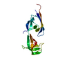 1eifSC S: Starting model for refinement C: citing same article ( |
|---|---|
| Similar structure data | |
| Other databases |


-Links
 PDBj
PDBj- Assembly
| Deposited unit | 
| ||||||||
|---|---|---|---|---|---|---|---|---|---|
| 1 |
| ||||||||
| Unit cell |
|
-Components
| #1: Protein | Mass: 14653.239 Da / Num. of mol.: 1 Source method: isolated from a genetically manipulated source Source: (gene. exp.) Methanocaldococcus jannaschii (archaea)Strain: DSM 2661 / Plasmid: PET21A/PSJS1240 / Species (production host): Escherichia coli / Production host:  |
|---|---|
| #2: Water | ChemComp-HOH /  Mass: 18.015 Da / Num. of mol.: 108 / Source method: isolated from a natural source / Formula: H2O Mass: 18.015 Da / Num. of mol.: 108 / Source method: isolated from a natural source / Formula: H2O |
-Experimental details
-Experiment
| Experiment | Method: X-RAY DIFFRACTION / Number of used crystals: 1 |
|---|
- Sample preparation
| Crystal | Density Matthews: 2.22 Å3/Da / Density % sol: 45 % | ||||||||||||||||||||||||||||||
|---|---|---|---|---|---|---|---|---|---|---|---|---|---|---|---|---|---|---|---|---|---|---|---|---|---|---|---|---|---|---|---|
| Crystal grow | pH: 8.5 / Details: 0.1M TRIS-HCL (PH 8.5), 0.2M MGCL2, 30% PEG4000 | ||||||||||||||||||||||||||||||
| Crystal | *PLUS | ||||||||||||||||||||||||||||||
| Crystal grow | *PLUS Temperature: 4 ℃ / Method: vapor diffusion | ||||||||||||||||||||||||||||||
| Components of the solutions | *PLUS
|
-Data collection
| Diffraction |
| ||||||||||||||||||||||||
|---|---|---|---|---|---|---|---|---|---|---|---|---|---|---|---|---|---|---|---|---|---|---|---|---|---|
| Diffraction source |
| ||||||||||||||||||||||||
| Detector | Type: RIGAKU RAXIS IIC / Detector: IMAGE PLATE / Date: Jul 1, 1997 / Details: MSC FOCUSING MIRROR | ||||||||||||||||||||||||
| Radiation | Protocol: SINGLE WAVELENGTH / Monochromatic (M) / Laue (L): M / Scattering type: x-ray | ||||||||||||||||||||||||
| Radiation wavelength | Wavelength: 1.54 Å / Relative weight: 1 | ||||||||||||||||||||||||
| Reflection | Resolution: 1.8→50 Å / Num. obs: 11659 / % possible obs: 96.7 % / Observed criterion σ(I): -3 / Redundancy: 1.98 % / Biso Wilson estimate: 22.1 Å2 / Rmerge(I) obs: 0.045 / Rsym value: 0.045 / Net I/σ(I): 16.2 | ||||||||||||||||||||||||
| Reflection shell | Resolution: 1.8→1.83 Å / Redundancy: 1.6 % / Rmerge(I) obs: 0.233 / Mean I/σ(I) obs: 3.7 / Rsym value: 0.233 / % possible all: 91.2 |

- Processing
| Software |
| ||||||||||||||||||||||||||||||||||||||||||||||||||||||||||||||||||||||||||||||||
|---|---|---|---|---|---|---|---|---|---|---|---|---|---|---|---|---|---|---|---|---|---|---|---|---|---|---|---|---|---|---|---|---|---|---|---|---|---|---|---|---|---|---|---|---|---|---|---|---|---|---|---|---|---|---|---|---|---|---|---|---|---|---|---|---|---|---|---|---|---|---|---|---|---|---|---|---|---|---|---|---|---|
| Refinement | Method to determine structure: MOLECULAR REPLACEMENT Starting model: 1EIF Resolution: 1.8→20 Å / Rfactor Rfree error: 0.007 / Data cutoff high rms absF: 1097804.99 / Isotropic thermal model: RESTRAINED / Cross valid method: THROUGHOUT / σ(F): 2
| ||||||||||||||||||||||||||||||||||||||||||||||||||||||||||||||||||||||||||||||||
| Solvent computation | Solvent model: FLAT MODEL / Bsol: 48.9 Å2 / ksol: 0.397 e/Å3 | ||||||||||||||||||||||||||||||||||||||||||||||||||||||||||||||||||||||||||||||||
| Displacement parameters | Biso mean: 25.3 Å2
| ||||||||||||||||||||||||||||||||||||||||||||||||||||||||||||||||||||||||||||||||
| Refine analyze |
| ||||||||||||||||||||||||||||||||||||||||||||||||||||||||||||||||||||||||||||||||
| Refinement step | Cycle: LAST / Resolution: 1.8→20 Å
| ||||||||||||||||||||||||||||||||||||||||||||||||||||||||||||||||||||||||||||||||
| Refine LS restraints |
| ||||||||||||||||||||||||||||||||||||||||||||||||||||||||||||||||||||||||||||||||
| LS refinement shell | Resolution: 1.8→1.91 Å / Rfactor Rfree error: 0.023 / Total num. of bins used: 6
| ||||||||||||||||||||||||||||||||||||||||||||||||||||||||||||||||||||||||||||||||
| Xplor file |
| ||||||||||||||||||||||||||||||||||||||||||||||||||||||||||||||||||||||||||||||||
| Software | *PLUS Name: CNS / Version: 0.3 / Classification: refinement | ||||||||||||||||||||||||||||||||||||||||||||||||||||||||||||||||||||||||||||||||
| Refinement | *PLUS Highest resolution: 1.8 Å / Lowest resolution: 20 Å / σ(F): 2 / % reflection Rfree: 10.3 % | ||||||||||||||||||||||||||||||||||||||||||||||||||||||||||||||||||||||||||||||||
| Solvent computation | *PLUS | ||||||||||||||||||||||||||||||||||||||||||||||||||||||||||||||||||||||||||||||||
| Displacement parameters | *PLUS Biso mean: 25.3 Å2 | ||||||||||||||||||||||||||||||||||||||||||||||||||||||||||||||||||||||||||||||||
| Refine LS restraints | *PLUS
| ||||||||||||||||||||||||||||||||||||||||||||||||||||||||||||||||||||||||||||||||
| LS refinement shell | *PLUS Rfactor Rfree: 0.342 / % reflection Rfree: 11.9 % / Rfactor Rwork: 0.285 |
