[English] 日本語
Yorodumi
- PDB-1bkb: INITIATION FACTOR 5A FROM ARCHEBACTERIUM PYROBACULUM AEROPHILUM -

+
Open data


ID or keywords:

Loading...

-
Basic information

Entry
Database: PDB / ID: 1bkb
TitleINITIATION FACTOR 5A FROM ARCHEBACTERIUM PYROBACULUM AEROPHILUM
ComponentsTRANSLATION INITIATION FACTOR 5A
KeywordsTRANSLATION / TRANSLATION INITIATION FACTOR
Function / homology
Function and homology information


positive regulation of translational termination / positive regulation of translational elongation / translation elongation factor activity / translation initiation factor activity / ribosome binding / cytoplasm
Similarity search - Function
Translation elongation factor IF5A, archaeal / Translation elongation factor, KOW-like / Elongation factor P (EF-P) KOW-like domain / Translation elongation factor, IF5A, hypusine site / Eukaryotic initiation factor 5A hypusine signature. / Eukaryotic elongation factor 5A hypusine, DNA-binding OB fold / Translation elongation factor IF5A-like / Translation elongation factor, IF5A C-terminal / Eukaryotic elongation factor 5A hypusine, DNA-binding OB fold / SH3 type barrels. - #30 ...Translation elongation factor IF5A, archaeal / Translation elongation factor, KOW-like / Elongation factor P (EF-P) KOW-like domain / Translation elongation factor, IF5A, hypusine site / Eukaryotic initiation factor 5A hypusine signature. / Eukaryotic elongation factor 5A hypusine, DNA-binding OB fold / Translation elongation factor IF5A-like / Translation elongation factor, IF5A C-terminal / Eukaryotic elongation factor 5A hypusine, DNA-binding OB fold / SH3 type barrels. - #30 / Nucleic acid-binding proteins / SH3 type barrels. / OB fold (Dihydrolipoamide Acetyltransferase, E2P) / Translation protein SH3-like domain superfamily / Ribosomal protein L2, domain 2 / Roll / Nucleic acid-binding, OB-fold / Beta Barrel / Mainly Beta
Similarity search - Domain/homology
Translation initiation factor 5A
Similarity search - Component
Biological speciesPyrobaculum aerophilum (archaea)
MethodX-RAY DIFFRACTION / SYNCHROTRON / MAD / Resolution: 1.75 Å
AuthorsPeat, T.S. / Newman, J. / Waldo, G.S. / Berendzen, J. / Terwilliger, T.C.
CitationJournal: Structure / Year: 1998
Title: Structure of translation initiation factor 5A from Pyrobaculum aerophilum at 1.75 A resolution.
Authors: Peat, T.S. / Newman, J. / Waldo, G.S. / Berendzen, J. / Terwilliger, T.C.
History
DepositionJul 5, 1998Processing site: BNL
Revision 1.0Nov 4, 1998Provider: repository / Type: Initial release
Revision 1.1Mar 24, 2008Group: Version format compliance
Revision 1.2Jul 13, 2011Group: Version format compliance

-
Structure visualization

Structure viewerMolecule:
MolmilJmol/JSmol

Downloads & links

-
Assembly

Deposited unit
A: TRANSLATION INITIATION FACTOR 5A


Theoretical massNumber of molelcules
Total (without water)15,3341
Polymers15,3341
Non-polymers00
Water2,576143
1
A: TRANSLATION INITIATION FACTOR 5A

A: TRANSLATION INITIATION FACTOR 5A

A: TRANSLATION INITIATION FACTOR 5A

A: TRANSLATION INITIATION FACTOR 5A


Theoretical massNumber of molelcules
Total (without water)61,3374
Polymers61,3374
Non-polymers00
Water724
TypeNameSymmetry operationNumber
identity operation1_555x,y,z1
crystal symmetry operation4_665y+1,-x+1,z1
crystal symmetry operation3_645-y+1,x-1,z1
crystal symmetry operation2_755-x+2,-y,z1
Unit cell
Length a, b, c (Å)114.130, 114.130, 32.590
Angle α, β, γ (deg.)90.00, 90.00, 90.00
Int Tables number79
Space group name H-MI4

-
Components

#1: Protein TRANSLATION INITIATION FACTOR 5A


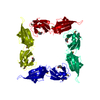
Mass: 15334.187 Da / Num. of mol.: 1 / Mutation: YES
Source method: isolated from a genetically manipulated source
Details: MSE HAS REPLACED MET, AS SELENO-METHIONINE WAS INCORPORATED INTO THE PROTEIN
Source: (gene. exp.) Pyrobaculum aerophilum (archaea) / Plasmid: PET / Species (production host): Escherichia coli / Production host: Escherichia coli BL21 (bacteria) / Strain (production host): BL21 / References: UniProt: P56635
#2: Water ChemComp-HOH / water / Water


Mass: 18.015 Da / Num. of mol.: 143 / Source method: isolated from a natural source / Formula: H2O

-
Experimental details

-
Experiment

ExperimentMethod: X-RAY DIFFRACTION / Number of used crystals: 1

-
Sample preparation

CrystalDensity Matthews: 3.53 Å3/Da / Density % sol: 60 %
Description: A THREE WAVELENGTH DATA SET WAS USED TO FIND THE SELENIUM POSITIONS AND PHASE THE ELECTRON DENSITY MAPS.
Crystal growTemperature: 281 K / pH: 7.5
Details: PROTEIN WAS CRYSTALLIZED AT 8 DEGREES IN 50MM HEPES PH 7.5, 6-8% PEG 4000, 5MM BETA- MERCAPTOETHANOL, temperature 281K
Crystal grow
*PLUS
Temperature: 8 ℃ / Method: unknown
Components of the solutions
*PLUS
IDConc.Common nameCrystal-IDSol-IDDetails
150 mMHEPES11
25-10 %PEG400011
35 mMdithiothreitol11or 5mM beta-mercaptoethanol

-
Data collection

DiffractionMean temperature: 110 K
Diffraction sourceSource: SYNCHROTRON / Site: NSLS / Beamline: X8C / Wavelength: 0.9788
DetectorType: MARRESEARCH / Detector: IMAGE PLATE / Date: May 1, 1998
RadiationMonochromator: SINGLE CRYSTAL / Protocol: SINGLE WAVELENGTH / Monochromatic (M) / Laue (L): M / Scattering type: x-ray
Radiation wavelengthWavelength: 0.9788 Å / Relative weight: 1
ReflectionResolution: 1.75→50 Å / Num. obs: 21532 / % possible obs: 99.8 % / Observed criterion σ(I): 0 / Redundancy: 7.8 % / Biso Wilson estimate: 17.1 Å2 / Rmerge(I) obs: 0.062 / Rsym value: 0.054 / Net I/σ(I): 8
Reflection shellResolution: 1.75→1.83 Å / Redundancy: 7.5 % / Rmerge(I) obs: 0.267 / Mean I/σ(I) obs: 3.3 / Rsym value: 0.23 / % possible all: 99.8
Reflection
*PLUS
Num. obs: 21537 / Redundancy: 7.9 % / Rmerge(I) obs: 0.053
Reflection shell
*PLUS
% possible obs: 99.3 % / Rmerge(I) obs: 0.23

-
Processing

Software
NameVersionClassification
CNS0.3refinement
SOLVEphasing
DENZOdata reduction
MOSFLMdata reduction
CCP4data scaling
CNS0.3phasing
RefinementMethod to determine structure: MAD / Resolution: 1.75→50 Å / Rfactor Rfree error: 0.007 / Isotropic thermal model: RESTRAINED / Cross valid method: THROUGHOUT / σ(F): 0
Details: ALTHOUGH AN ANOMALOUS PARAMETER FILE SHOULD HAVE BEEN USED IN REFINEMENT (SE ATOMS IN STRUCTURE), THIS WAS NOT DONE. DATA CUTOFF HIGH (ABS(F)) : 1392611.86 DATA CUTOFF LOW (ABS(F)) : 0.00
RfactorNum. reflection% reflectionSelection details
Rfree0.236 1052 4.9 %RANDOM
Rwork0.214 ---
obs0.214 21532 99.7 %-
Solvent computationSolvent model: COMBINATION / Bsol: 110.6 Å2 / ksol: 0.37 e/Å3
Displacement parametersBiso mean: 24.3 Å2
Baniso -1Baniso -2Baniso -3
1-3.2 Å20 Å20 Å2
2--3.2 Å20 Å2
3----6.4 Å2
Refine analyze
FreeObs
Luzzati coordinate error0.23 Å0.21 Å
Luzzati d res low-5 Å
Luzzati sigma a0.15 Å0.13 Å
Refinement stepCycle: LAST / Resolution: 1.75→50 Å
ProteinNucleic acidLigandSolventTotal
Num. atoms1033 0 0 143 1176
Refine LS restraints
Refine-IDTypeDev idealDev ideal target
X-RAY DIFFRACTIONc_bond_d0.005
X-RAY DIFFRACTIONc_bond_d_na
X-RAY DIFFRACTIONc_bond_d_prot
X-RAY DIFFRACTIONc_angle_d
X-RAY DIFFRACTIONc_angle_d_na
X-RAY DIFFRACTIONc_angle_d_prot
X-RAY DIFFRACTIONc_angle_deg1.3
X-RAY DIFFRACTIONc_angle_deg_na
X-RAY DIFFRACTIONc_angle_deg_prot
X-RAY DIFFRACTIONc_dihedral_angle_d25.5
X-RAY DIFFRACTIONc_dihedral_angle_d_na
X-RAY DIFFRACTIONc_dihedral_angle_d_prot
X-RAY DIFFRACTIONc_improper_angle_d0.7
X-RAY DIFFRACTIONc_improper_angle_d_na
X-RAY DIFFRACTIONc_improper_angle_d_prot
X-RAY DIFFRACTIONc_mcbond_it0.641.5
X-RAY DIFFRACTIONc_mcangle_it1.132
X-RAY DIFFRACTIONc_scbond_it0.412
X-RAY DIFFRACTIONc_scangle_it0.732.5
LS refinement shellResolution: 1.75→1.83 Å / Rfactor Rfree error: 0.022 / Total num. of bins used: 8
RfactorNum. reflection% reflection
Rfree0.278 153 5.8 %
Rwork0.253 2507 -
obs--99.9 %
Xplor file
Refine-IDSerial noParam fileTopol file
X-RAY DIFFRACTION1PROTEIN_REP.PARAMPROTEIN.TOP
X-RAY DIFFRACTION2WATER_REP.PARAMWATER.TOP
Software
*PLUS
Name: CNS / Version: 0.3 / Classification: refinement
Refine LS restraints
*PLUS
Refine-IDTypeDev ideal
X-RAY DIFFRACTIONc_dihedral_angle_d
X-RAY DIFFRACTIONc_dihedral_angle_deg25.5
X-RAY DIFFRACTIONc_improper_angle_d
X-RAY DIFFRACTIONc_improper_angle_deg0.7

+
About Yorodumi

-
News

-
Feb 9, 2022. New format data for meta-information of EMDB entries

New format data for meta-information of EMDB entries

  • Version 3 of the EMDB header file is now the official format.
  • The previous official version 1.9 will be removed from the archive.

Related info.:EMDB header

External links:wwPDB to switch to version 3 of the EMDB data model

-
Aug 12, 2020. Covid-19 info

Covid-19 info

URL: https://pdbj.org/emnavi/covid19.php

New page: Covid-19 featured information page in EM Navigator.

Related info.:Covid-19 info / Mar 5, 2020. Novel coronavirus structure data

+
Mar 5, 2020. Novel coronavirus structure data

Novel coronavirus structure data

Related info.:Yorodumi Speices / Aug 12, 2020. Covid-19 info

External links:COVID-19 featured content - PDBj / Molecule of the Month (242):Coronavirus Proteases

+
Jan 31, 2019. EMDB accession codes are about to change! (news from PDBe EMDB page)

EMDB accession codes are about to change! (news from PDBe EMDB page)

  • The allocation of 4 digits for EMDB accession codes will soon come to an end. Whilst these codes will remain in use, new EMDB accession codes will include an additional digit and will expand incrementally as the available range of codes is exhausted. The current 4-digit format prefixed with “EMD-” (i.e. EMD-XXXX) will advance to a 5-digit format (i.e. EMD-XXXXX), and so on. It is currently estimated that the 4-digit codes will be depleted around Spring 2019, at which point the 5-digit format will come into force.
  • The EM Navigator/Yorodumi systems omit the EMD- prefix.

Related info.:Q: What is EMD? / ID/Accession-code notation in Yorodumi/EM Navigator

External links:EMDB Accession Codes are Changing Soon! / Contact to PDBj

+
Jul 12, 2017. Major update of PDB

Major update of PDB

  • wwPDB released updated PDB data conforming to the new PDBx/mmCIF dictionary.
  • This is a major update changing the version number from 4 to 5, and with Remediation, in which all the entries are updated.
  • In this update, many items about electron microscopy experimental information are reorganized (e.g. em_software).
  • Now, EM Navigator and Yorodumi are based on the updated data.

External links:wwPDB Remediation / Enriched Model Files Conforming to OneDep Data Standards Now Available in the PDB FTP Archive

-
Yorodumi

Thousand views of thousand structures

  • Yorodumi is a browser for structure data from EMDB, PDB, SASBDB, etc.
  • This page is also the successor to EM Navigator detail page, and also detail information page/front-end page for Omokage search.
  • The word "yorodu" (or yorozu) is an old Japanese word meaning "ten thousand". "mi" (miru) is to see.

Related info.:EMDB / PDB / SASBDB / Comparison of 3 databanks / Yorodumi Search / Aug 31, 2016. New EM Navigator & Yorodumi / Yorodumi Papers / Jmol/JSmol / Function and homology information / Changes in new EM Navigator and Yorodumi

Read more