Resolution: 2.48→2.63 Å / Mean I/σ(I) obs: 3.97 / Num. unique all: 1885 / % possible all: 83.8
-
Processing
Software
Name
Version
Classification
MAR345dtb
datacollection
PHASER
phasing
BUSTER
2.9.1
refinement
XDS
datareduction
XDS
datascaling
Refinement
Method to determine structure: MOLECULAR REPLACEMENT / Resolution: 2.48→20.86 Å / SU R Cruickshank DPI: 0.873 / Cross valid method: THROUGHOUT / σ(F): 0
Rfactor
Num. reflection
% reflection
Selection details
Rfree
0.2499
659
5 %
RANDOM
Rwork
0.194
-
-
-
all
0.1967
-
-
-
obs
0.1967
13169
93.9 %
-
Displacement parameters
Biso mean: 43.35 Å2
Refine analyze
Luzzati coordinate error obs: 0.318 Å
Refinement step
Cycle: LAST / Resolution: 2.48→20.86 Å
Protein
Nucleic acid
Ligand
Solvent
Total
Num. atoms
2923
0
0
129
3052
Refine LS restraints
Refine-ID
Type
Dev ideal
Number
Weight
X-RAY DIFFRACTION
t_bond_d
0.01
2947
2
X-RAY DIFFRACTION
t_angle_deg
1.17
3975
2
X-RAY DIFFRACTION
t_dihedral_angle_d
1079
2
X-RAY DIFFRACTION
t_trig_c_planes
80
2
X-RAY DIFFRACTION
t_gen_planes
423
5
X-RAY DIFFRACTION
t_it
2947
20
X-RAY DIFFRACTION
t_omega_torsion
2.75
X-RAY DIFFRACTION
t_other_torsion
18
X-RAY DIFFRACTION
t_chiral_improper_torsion
405
5
X-RAY DIFFRACTION
t_ideal_dist_contact
3483
4
LS refinement shell
Resolution: 2.48→2.68 Å / Total num. of bins used: 7
Rfactor
Num. reflection
% reflection
Rfree
0.3135
125
5.01 %
Rwork
0.2155
2368
-
all
0.2202
2493
-
obs
-
-
93.9 %
+
About Yorodumi
-
News
-
Feb 9, 2022. New format data for meta-information of EMDB entries
New format data for meta-information of EMDB entries
Version 3 of the EMDB header file is now the official format.
The previous official version 1.9 will be removed from the archive.
In the structure databanks used in Yorodumi, some data are registered as the other names, "COVID-19 virus" and "2019-nCoV". Here are the details of the virus and the list of structure data.
Jan 31, 2019. EMDB accession codes are about to change! (news from PDBe EMDB page)
EMDB accession codes are about to change! (news from PDBe EMDB page)
The allocation of 4 digits for EMDB accession codes will soon come to an end. Whilst these codes will remain in use, new EMDB accession codes will include an additional digit and will expand incrementally as the available range of codes is exhausted. The current 4-digit format prefixed with “EMD-” (i.e. EMD-XXXX) will advance to a 5-digit format (i.e. EMD-XXXXX), and so on. It is currently estimated that the 4-digit codes will be depleted around Spring 2019, at which point the 5-digit format will come into force.
The EM Navigator/Yorodumi systems omit the EMD- prefix.
Related info.:Q: What is EMD? / ID/Accession-code notation in Yorodumi/EM Navigator
Yorodumi is a browser for structure data from EMDB, PDB, SASBDB, etc.
This page is also the successor to EM Navigator detail page, and also detail information page/front-end page for Omokage search.
The word "yorodu" (or yorozu) is an old Japanese word meaning "ten thousand". "mi" (miru) is to see.
Related info.:EMDB / PDB / SASBDB / Comparison of 3 databanks / Yorodumi Search / Aug 31, 2016. New EM Navigator & Yorodumi / Yorodumi Papers / Jmol/JSmol / Function and homology information / Changes in new EM Navigator and Yorodumi
 Movie
Movie Controller

 Yorodumi
 Open data
 Basic information
 Components
 Keywords
 Function and homology information
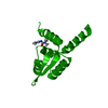
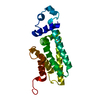
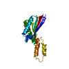
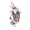
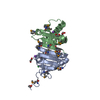
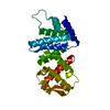
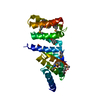
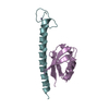
 Xylella fastidiosa (bacteria)
 X-RAY DIFFRACTION /
  Authors
 Citation
 Structure visualization
 Downloads & links
 Other downloads

 PDBj
PDBj Assembly
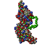
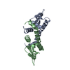
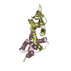
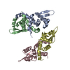
 Mass: 18.015 Da / Num. of mol.: 129 / Source method: isolated from a natural source / Formula: H2O
 Sample preparation
 / Beamline: W01B-MX2 / Wavelength: 1.4333 Å
 Processing