 Movie
Movie Controller


+ Open data

- Basic information
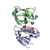
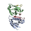
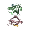
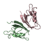
| Entry | Database: PDB / ID: 2ego | ||||||
|---|---|---|---|---|---|---|---|
| Title | Crystal Structure of Tamalin PDZ Domain | ||||||
 Components Components | General receptor for phosphoinositides 1-associated scaffold protein | ||||||
 Keywords Keywords | PROTEIN BINDING / PDZ domain / ligand-free | ||||||
| Function / homology |  Function and homology information Function and homology informationregulation of postsynaptic neurotransmitter receptor internalization / PDZ domain binding / small GTPase binding / postsynaptic membrane / postsynaptic density / perinuclear region of cytoplasm / glutamatergic synapse / signal transduction / identical protein binding / plasma membrane Similarity search - Function | ||||||
| Biological species |  | ||||||
| Method |  X-RAY DIFFRACTION / SYNCHROTRON / MOLECULAR REPLACEMENT / Resolution: 1.8 Å X-RAY DIFFRACTION / SYNCHROTRON / MOLECULAR REPLACEMENT / Resolution: 1.8 Å | ||||||
 Authors Authors | Sugi, T. / Oyama, T. / Muto, T. / Nakanishi, S. / Morikawa, K. / Jingami, H. | ||||||
 Citation Citation | Journal: Embo J. / Year: 2007 Title: Crystal structures of autoinhibitory PDZ domain of Tamalin: implications for metabotropic glutamate receptor trafficking regulation Authors: Sugi, T. / Oyama, T. / Muto, T. / Nakanishi, S. / Morikawa, K. / Jingami, H. | ||||||
| History |
|
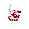
- Structure visualization
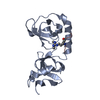
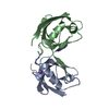
| Structure viewer | Molecule: MolmilJmol/JSmol |
|---|
- Downloads & links
-Download
| PDBx/mmCIF format | 2ego.cif.gz | 47.7 KB | Display | PDBx/mmCIF format |
|---|---|---|---|---|
| PDB format | pdb2ego.ent.gz | 33.8 KB | Display | PDB format |
| PDBx/mmJSON format | 2ego.json.gz | Tree view | PDBx/mmJSON format | |
| Others |  Other downloads Other downloads |
-Validation report
| Arichive directory | https://data.pdbj.org/pub/pdb/validation_reports/eg/2egoftp://data.pdbj.org/pub/pdb/validation_reports/eg/2ego | HTTPS FTP |
|---|
-Related structure data
| Related structure data |  2egkC  2egnC 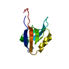 1g9oS C: citing same article ( S: Starting model for refinement |
|---|---|
| Similar structure data |

-Links
 PDBj
PDBj- Assembly
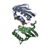
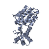
| Deposited unit | 
| ||||||||
|---|---|---|---|---|---|---|---|---|---|
| 1 |
| ||||||||
| Unit cell |
|
-Components
| #1: Protein | Mass: 10643.935 Da / Num. of mol.: 2 / Fragment: PDZ domain / Mutation: C135A Source method: isolated from a genetically manipulated source Source: (gene. exp.)  #2: Water | ChemComp-HOH / |  Mass: 18.015 Da / Num. of mol.: 107 / Source method: isolated from a natural source / Formula: H2O Mass: 18.015 Da / Num. of mol.: 107 / Source method: isolated from a natural source / Formula: H2O |
|---|
-Experimental details
-Experiment
| Experiment | Method: X-RAY DIFFRACTION / Number of used crystals: 1 |
|---|
- Sample preparation
| Crystal | Density Matthews: 2.32 Å3/Da / Density % sol: 47.08 % |
|---|---|
| Crystal grow | Temperature: 293 K / Method: vapor diffusion, hanging drop / pH: 7.2 Details: 20% PEG 3350, 0.2 M potassium fluoride, pH 7.2, VAPOR DIFFUSION, HANGING DROP, temperature 293K |
-Data collection
| Diffraction | Mean temperature: 298 K |
|---|---|
| Diffraction source | Source: SYNCHROTRON / Site: Photon Factory  / Beamline: BL-6B / Wavelength: 1 Å / Beamline: BL-6B / Wavelength: 1 Å |
| Detector | Type: RIGAKU RAXIS IV / Detector: IMAGE PLATE / Date: Nov 12, 2006 |
| Radiation | Protocol: SINGLE WAVELENGTH / Monochromatic (M) / Laue (L): M / Scattering type: x-ray |
| Radiation wavelength | Wavelength: 1 Å / Relative weight: 1 |
| Reflection | Resolution: 1.8→50 Å / Num. all: 18424 / Num. obs: 18300 / % possible obs: 99.3 % / Observed criterion σ(F): 0 / Redundancy: 3.6 % / Biso Wilson estimate: -1 Å2 / Rmerge(I) obs: 0.091 / Net I/σ(I): 7.2 |
| Reflection shell | Resolution: 1.8→1.87 Å / Redundancy: 3.6 % / Rmerge(I) obs: 0.276 / % possible all: 97.1 |
- Processing
| Software |
| ||||||||||||||||||||||||||||||||||||||||||||||||||||||||||||||||||||||||||||||||
|---|---|---|---|---|---|---|---|---|---|---|---|---|---|---|---|---|---|---|---|---|---|---|---|---|---|---|---|---|---|---|---|---|---|---|---|---|---|---|---|---|---|---|---|---|---|---|---|---|---|---|---|---|---|---|---|---|---|---|---|---|---|---|---|---|---|---|---|---|---|---|---|---|---|---|---|---|---|---|---|---|---|
| Refinement | Method to determine structure: MOLECULAR REPLACEMENT Starting model: PDB ENTRY 1G9O Resolution: 1.8→41.14 Å / Rfactor Rfree error: 0.034 / Data cutoff high absF: 233285.59 / Data cutoff low absF: 0 / Isotropic thermal model: RESTRAINED / Cross valid method: THROUGHOUT / σ(F): 0
| ||||||||||||||||||||||||||||||||||||||||||||||||||||||||||||||||||||||||||||||||
| Solvent computation | Solvent model: FLAT MODEL / Bsol: 10 Å2 / ksol: 0.421173 e/Å3 | ||||||||||||||||||||||||||||||||||||||||||||||||||||||||||||||||||||||||||||||||
| Displacement parameters | Biso mean: 14 Å2
| ||||||||||||||||||||||||||||||||||||||||||||||||||||||||||||||||||||||||||||||||
| Refine analyze |
| ||||||||||||||||||||||||||||||||||||||||||||||||||||||||||||||||||||||||||||||||
| Refinement step | Cycle: LAST / Resolution: 1.8→41.14 Å
| ||||||||||||||||||||||||||||||||||||||||||||||||||||||||||||||||||||||||||||||||
| Refine LS restraints |
| ||||||||||||||||||||||||||||||||||||||||||||||||||||||||||||||||||||||||||||||||
| LS refinement shell | Resolution: 1.8→1.91 Å / Rfactor Rfree error: 0.135 / Total num. of bins used: 6
| ||||||||||||||||||||||||||||||||||||||||||||||||||||||||||||||||||||||||||||||||
| Xplor file |
|
