 Movie
Movie Controller

[English] 日本語
 Yorodumi
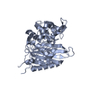
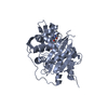
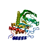
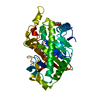
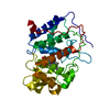
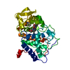
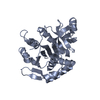
- PDB-2ecu: Crystal structure of flavin reductase component (HpaC) of 4-hydro... -
+ Open data

- Basic information
| Entry | Database: PDB / ID: 2ecu | ||||||
|---|---|---|---|---|---|---|---|
| Title | Crystal structure of flavin reductase component (HpaC) of 4-hydroxyphenylacetate 3-monooxygenase | ||||||
 Components Components | flavin reductase (HpaC) of 4-hydroxyphenylacetate 3-monooxygnease | ||||||
 Keywords Keywords | OXIDOREDUCTASE / flavin reductase / flavin diffusible / two-component monooxygenase | ||||||
| Function / homology |  Function and homology information Function and homology informationflavin reductase (NADH) / pyrimidine nucleobase catabolic process / flavin reductase (NADH) activity / riboflavin reductase (NADPH) activity / FMN binding Similarity search - Function | ||||||
| Biological species |   Thermus thermophilus (bacteria) Thermus thermophilus (bacteria) | ||||||
| Method |  X-RAY DIFFRACTION / SYNCHROTRON / MOLECULAR REPLACEMENT / Resolution: 1.3 Å X-RAY DIFFRACTION / SYNCHROTRON / MOLECULAR REPLACEMENT / Resolution: 1.3 Å | ||||||
 Authors Authors | Kim, S.H. / Hisano, T. / Iwasaki, W. / Ebihara, A. / Miki, K. | ||||||
 Citation Citation | Journal: Proteins / Year: 2008 Title: Crystal structure of the flavin reductase component (HpaC) of 4-hydroxyphenylacetate 3-monooxygenase from Thermus thermophilus HB8: Structural basis for the flavin affinity Authors: Kim, S.H. / Hisano, T. / Iwasaki, W. / Ebihara, A. / Miki, K. | ||||||
| History |
|
- Structure visualization
| Structure viewer | Molecule: MolmilJmol/JSmol |
|---|
- Downloads & links
-Download
| PDBx/mmCIF format | 2ecu.cif.gz | 79.3 KB | Display | PDBx/mmCIF format |
|---|---|---|---|---|
| PDB format | pdb2ecu.ent.gz | 58.5 KB | Display | PDB format |
| PDBx/mmJSON format | 2ecu.json.gz | Tree view | PDBx/mmJSON format | |
| Others |  Other downloads Other downloads |
-Validation report
| Summary document | 2ecu_validation.pdf.gz | 915.2 KB | Display | wwPDB validaton report |
|---|---|---|---|---|
| Full document | 2ecu_full_validation.pdf.gz | 912.9 KB | Display | |
| Data in XML | 2ecu_validation.xml.gz | 17.4 KB | Display | |
| Data in CIF | 2ecu_validation.cif.gz | 26.1 KB | Display | |
| Arichive directory | https://data.pdbj.org/pub/pdb/validation_reports/ec/2ecuftp://data.pdbj.org/pub/pdb/validation_reports/ec/2ecu | HTTPS FTP |
-Related structure data


-Links
 PDBj
PDBj- Assembly
| Deposited unit | 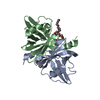
| ||||||||
|---|---|---|---|---|---|---|---|---|---|
| 1 |
| ||||||||
| Unit cell |
|
-Components
| #1: Protein | Mass: 16059.578 Da / Num. of mol.: 2 / Mutation: E131G Source method: isolated from a genetically manipulated source Source: (gene. exp.) Thermus thermophilus (bacteria) / Strain: HB8 / Gene: TTHA0961 / Plasmid: pET11a / Species (production host): Escherichia coli / Production host: #2: Chemical | ChemComp-1PG / | 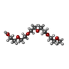  Mass: 252.305 Da / Num. of mol.: 1 / Source method: obtained synthetically / Formula: C11H24O6 Mass: 252.305 Da / Num. of mol.: 1 / Source method: obtained synthetically / Formula: C11H24O6#3: Chemical | ChemComp-12P / |   Mass: 546.646 Da / Num. of mol.: 1 / Source method: obtained synthetically / Formula: C24H50O13 / Comment: precipitant*YM Mass: 546.646 Da / Num. of mol.: 1 / Source method: obtained synthetically / Formula: C24H50O13 / Comment: precipitant*YM#4: Water | ChemComp-HOH / |  Mass: 18.015 Da / Num. of mol.: 400 / Source method: isolated from a natural source / Formula: H2O Mass: 18.015 Da / Num. of mol.: 400 / Source method: isolated from a natural source / Formula: H2ONonpolymer details | PEG1000 AND PEG8000 ARE USED IN THE CRYSTALLIZATION CONDITION OF THIS PROTEIN. THEREFORE IT IS ...PEG1000 AND PEG8000 ARE USED IN THE CRYSTALLIZ | |
|---|
-Experimental details
-Experiment
| Experiment | Method: X-RAY DIFFRACTION / Number of used crystals: 1 |
|---|
- Sample preparation
| Crystal | Density Matthews: 2.11 Å3/Da / Density % sol: 41.69 % |
|---|---|
| Crystal grow | Temperature: 293 K / Method: vapor diffusion, hanging drop Details: 20% PEG 1000, 10% PEG 8000, 10% glycerol, VAPOR DIFFUSION, HANGING DROP, temperature 293K |
-Data collection
| Diffraction | Mean temperature: 100 K |
|---|---|
| Diffraction source | Source: SYNCHROTRON / Site: SPring-8  / Beamline: BL45XU / Wavelength: 1.02 Å / Beamline: BL45XU / Wavelength: 1.02 Å |
| Detector | Type: RIGAKU RAXIS V / Detector: IMAGE PLATE / Date: Apr 20, 2001 / Details: mirrors |
| Radiation | Monochromator: DIAMOND / Protocol: SINGLE WAVELENGTH / Monochromatic (M) / Laue (L): M / Scattering type: x-ray |
| Radiation wavelength | Wavelength: 1.02 Å / Relative weight: 1 |
| Reflection | Resolution: 1.3→41.53 Å / Num. obs: 62285 / % possible obs: 94.6 % / Observed criterion σ(I): 0 / Redundancy: 2.6 % / Biso Wilson estimate: 9.649 Å2 / Rmerge(I) obs: 0.062 / Net I/σ(I): 19.9 |
| Reflection shell | Resolution: 1.3→1.35 Å / Redundancy: 2.2 % / Rmerge(I) obs: 0.195 / Mean I/σ(I) obs: 8.24 / Num. unique all: 5450 / % possible all: 83.3 |
- Processing
| Software |
| ||||||||||||||||||||||||||||||||||||||||||
|---|---|---|---|---|---|---|---|---|---|---|---|---|---|---|---|---|---|---|---|---|---|---|---|---|---|---|---|---|---|---|---|---|---|---|---|---|---|---|---|---|---|---|---|
| Refinement | Method to determine structure: MOLECULAR REPLACEMENT Starting model: Se-derivatazed HpaC MAD Resolution: 1.3→41.53 Å / Isotropic thermal model: isotropic / Cross valid method: THROUGHOUT / σ(I): 0 / Stereochemistry target values: Engh & Huber
| ||||||||||||||||||||||||||||||||||||||||||
| Displacement parameters | Biso mean: 11.4 Å2
| ||||||||||||||||||||||||||||||||||||||||||
| Refine analyze |
| ||||||||||||||||||||||||||||||||||||||||||
| Refinement step | Cycle: LAST / Resolution: 1.3→41.53 Å
| ||||||||||||||||||||||||||||||||||||||||||
| Refine LS restraints |
| ||||||||||||||||||||||||||||||||||||||||||
| LS refinement shell | Refine-ID: X-RAY DIFFRACTION / Total num. of bins used: 6
|
