 Movie
Movie Controller

[English] 日本語
 Yorodumi
- PDB-2ecr: Crystal structure of the ligand-free form of the flavin reductase... -
+ Open data

- Basic information
| Entry | Database: PDB / ID: 2ecr | ||||||
|---|---|---|---|---|---|---|---|
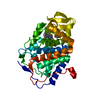
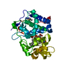
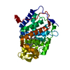
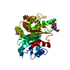
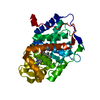
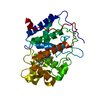
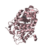
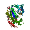
| Title | Crystal structure of the ligand-free form of the flavin reductase component (HpaC) of 4-hydroxyphenylacetate 3-monooxygenase | ||||||
 Components Components | flavin reductase component (HpaC) of 4-hydroxyphenylacetate 3-monooxygenase | ||||||
 Keywords Keywords | OXIDOREDUCTASE / Flavin reductase / flavin diffusible / two-component monooxygenase | ||||||
| Function / homology |  Function and homology information Function and homology informationflavin reductase (NADH) / pyrimidine nucleobase catabolic process / flavin reductase (NADH) activity / riboflavin reductase (NADPH) activity / FMN binding Similarity search - Function | ||||||
| Biological species |   Thermus thermophilus (bacteria) Thermus thermophilus (bacteria) | ||||||
| Method |  X-RAY DIFFRACTION / SYNCHROTRON / MOLECULAR REPLACEMENT / Resolution: 1.6 Å X-RAY DIFFRACTION / SYNCHROTRON / MOLECULAR REPLACEMENT / Resolution: 1.6 Å | ||||||
 Authors Authors | Kim, S.H. / Hisano, T. / Iwasaki, W. / Ebihara, A. / Miki, K. | ||||||
 Citation Citation | Journal: Proteins / Year: 2008 Title: Crystal structure of the flavin reductase component (HpaC) of 4-hydroxyphenylacetate 3-monooxygenase from Thermus thermophilus HB8: Structural basis for the flavin affinity Authors: Kim, S.H. / Hisano, T. / Iwasaki, W. / Ebihara, A. / Miki, K. #1: Journal: ACTA CRYSTALLOGR.,SECT.D / Year: 2003 Title: Crystallization and preliminary X-ray analysis of the small component of 4-hydroxyphenylacetate 3-monooxygenase (HpaC) and its cofactor complex from Thermus thermophilus HB8 Authors: Kim, S.H. / Miyatake, H. / Hisano, T. / Ohtani, N. / Miki, K. | ||||||
| History |
|
- Structure visualization
| Structure viewer | Molecule: MolmilJmol/JSmol |
|---|
- Downloads & links
-Download
| PDBx/mmCIF format | 2ecr.cif.gz | 72.7 KB | Display | PDBx/mmCIF format |
|---|---|---|---|---|
| PDB format | pdb2ecr.ent.gz | 55 KB | Display | PDB format |
| PDBx/mmJSON format | 2ecr.json.gz | Tree view | PDBx/mmJSON format | |
| Others |  Other downloads Other downloads |
-Validation report
| Arichive directory | https://data.pdbj.org/pub/pdb/validation_reports/ec/2ecrftp://data.pdbj.org/pub/pdb/validation_reports/ec/2ecr | HTTPS FTP |
|---|
-Related structure data


-Links
 PDBj
PDBj- Assembly
| Deposited unit | 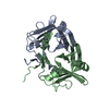
| ||||||||
|---|---|---|---|---|---|---|---|---|---|
| 1 |
| ||||||||
| Unit cell |
|
-Components
| #1: Protein | Mass: 16059.578 Da / Num. of mol.: 2 / Mutation: E131G Source method: isolated from a genetically manipulated source Source: (gene. exp.) Thermus thermophilus (bacteria) / Strain: HB8 / Gene: TTHA0961 / Plasmid: pET11a / Species (production host): Escherichia coli / Production host: #2: Water | ChemComp-HOH / |  Mass: 18.015 Da / Num. of mol.: 304 / Source method: isolated from a natural source / Formula: H2O Mass: 18.015 Da / Num. of mol.: 304 / Source method: isolated from a natural source / Formula: H2O |
|---|
-Experimental details
-Experiment
| Experiment | Method: X-RAY DIFFRACTION / Number of used crystals: 1 |
|---|
- Sample preparation
| Crystal | Density Matthews: 2.12 Å3/Da / Density % sol: 42.1 % |
|---|---|
| Crystal grow | Temperature: 293 K / Method: vapor diffusion, hanging drop / pH: 7 Details: 40% PEG 4000, 5% isopropyl alcohol, 0.1M HEPES-NaOH, 10% glycerol, pH 7.0, VAPOR DIFFUSION, HANGING DROP, temperature 293K |
-Data collection
| Diffraction | Mean temperature: 100 K |
|---|---|
| Diffraction source | Source: SYNCHROTRON / Site: SPring-8  / Beamline: BL45XU / Wavelength: 1 Å / Beamline: BL45XU / Wavelength: 1 Å |
| Detector | Type: RIGAKU RAXIS V / Detector: IMAGE PLATE / Date: Nov 27, 2002 / Details: mirrors |
| Radiation | Monochromator: DIAMOND / Protocol: SINGLE WAVELENGTH / Monochromatic (M) / Laue (L): M / Scattering type: x-ray |
| Radiation wavelength | Wavelength: 1 Å / Relative weight: 1 |
| Reflection | Resolution: 1.6→50 Å / Num. obs: 35392 / % possible obs: 99.4 % / Observed criterion σ(I): 0 / Redundancy: 2.8 % / Biso Wilson estimate: 13.466 Å2 / Rmerge(I) obs: 0.071 / Net I/σ(I): 18.7 |
| Reflection shell | Resolution: 1.6→1.66 Å / Redundancy: 3.6 % / Rmerge(I) obs: 0.314 / Mean I/σ(I) obs: 7.2 / Num. unique all: 3505 / % possible all: 99.7 |
- Processing
| Software |
| |||||||||||||||||||||||||||||||||||||||||||||||||
|---|---|---|---|---|---|---|---|---|---|---|---|---|---|---|---|---|---|---|---|---|---|---|---|---|---|---|---|---|---|---|---|---|---|---|---|---|---|---|---|---|---|---|---|---|---|---|---|---|---|---|
| Refinement | Method to determine structure: MOLECULAR REPLACEMENT Starting model: Selenomethionine-labeled crystal was used for phase determination, and the model was used for initial target model. Resolution: 1.6→41.4 Å / Isotropic thermal model: isotropic / Cross valid method: THROUGHOUT / σ(I): 0 / Stereochemistry target values: Engh & Huber
| |||||||||||||||||||||||||||||||||||||||||||||||||
| Displacement parameters | Biso mean: 15 Å2
| |||||||||||||||||||||||||||||||||||||||||||||||||
| Refine analyze |
| |||||||||||||||||||||||||||||||||||||||||||||||||
| Refinement step | Cycle: LAST / Resolution: 1.6→41.4 Å
| |||||||||||||||||||||||||||||||||||||||||||||||||
| Refine LS restraints |
| |||||||||||||||||||||||||||||||||||||||||||||||||
| LS refinement shell |
|
