 Movie
Movie Controller


+ Open data

- Basic information
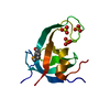
| Entry | Database: PDB / ID: 2ea9 | ||||||
|---|---|---|---|---|---|---|---|
| Title | Crystal structure of a hypothetical protein JW2626 from E.coli | ||||||
 Components Components | Hypothetical protein yfjZ | ||||||
 Keywords Keywords | STRUCTURAL GENOMICS / UNKNOWN FUNCTION / Escherichia coli / JW2626 / hypothetical protein / NPPSFA / National Project on Protein Structural and Functional Analyses / RIKEN Structural Genomics/Proteomics Initiative / RSGI | ||||||
| Function / homology | Antitoxin CbeA / Antitoxin CbeA superfamily / CbeA_antitoxin, type IV, cytoskeleton bundling-enhancing factor A / positive regulation of cytoskeleton organization / PAS domain / Beta-Lactamase / 2-Layer Sandwich / Alpha Beta / Antitoxin YfjZ Function and homology information Function and homology information | ||||||
| Biological species |  | ||||||
| Method |  X-RAY DIFFRACTION / SYNCHROTRON / MOLECULAR REPLACEMENT / Resolution: 2.1 Å X-RAY DIFFRACTION / SYNCHROTRON / MOLECULAR REPLACEMENT / Resolution: 2.1 Å | ||||||
 Authors Authors | Okazaki, N. / Kuramitsu, S. / Yamamoto, M. / Yokoyama, S. / RIKEN Structural Genomics/Proteomics Initiative (RSGI) | ||||||
 Citation Citation | Journal: To be Published Title: Crystal structure of a hypothetical protein JW2626 from E.coli Authors: Okazaki, N. / Kuramitsu, S. / Yamamoto, M. / Yokoyama, S. | ||||||
| History |
|
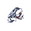
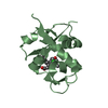
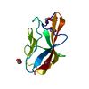
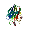
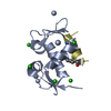
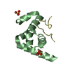
- Structure visualization
| Structure viewer | Molecule: MolmilJmol/JSmol |
|---|
- Downloads & links
-Download
| PDBx/mmCIF format | 2ea9.cif.gz | 33.9 KB | Display | PDBx/mmCIF format |
|---|---|---|---|---|
| PDB format | pdb2ea9.ent.gz | 21.7 KB | Display | PDB format |
| PDBx/mmJSON format | 2ea9.json.gz | Tree view | PDBx/mmJSON format | |
| Others |  Other downloads Other downloads |
-Validation report
| Summary document | 2ea9_validation.pdf.gz | 411.4 KB | Display | wwPDB validaton report |
|---|---|---|---|---|
| Full document | 2ea9_full_validation.pdf.gz | 414.3 KB | Display | |
| Data in XML | 2ea9_validation.xml.gz | 7.1 KB | Display | |
| Data in CIF | 2ea9_validation.cif.gz | 9 KB | Display | |
| Arichive directory | https://data.pdbj.org/pub/pdb/validation_reports/ea/2ea9ftp://data.pdbj.org/pub/pdb/validation_reports/ea/2ea9 | HTTPS FTP |
-Related structure data
| Related structure data |  2h28S S: Starting model for refinement |
|---|---|
| Similar structure data | |
| Other databases |
-Links
 PDBj
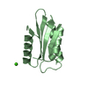
PDBj- Assembly
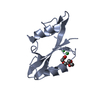
| Deposited unit | 
| ||||||||
|---|---|---|---|---|---|---|---|---|---|
| 1 |
| ||||||||
| Unit cell |
| ||||||||
| Details | The biological assembly is a monomer. |
-Components
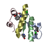
| #1: Protein | Mass: 11749.244 Da / Num. of mol.: 1 Source method: isolated from a genetically manipulated source Source: (gene. exp.) |
|---|---|
| #2: Water | ChemComp-HOH /  Mass: 18.015 Da / Num. of mol.: 64 / Source method: isolated from a natural source / Formula: H2O Mass: 18.015 Da / Num. of mol.: 64 / Source method: isolated from a natural source / Formula: H2O |
-Experimental details
-Experiment
| Experiment | Method: X-RAY DIFFRACTION / Number of used crystals: 1 |
|---|
- Sample preparation
| Crystal | Density Matthews: 2.35 Å3/Da / Density % sol: 47.74 % |
|---|---|
| Crystal grow | Temperature: 291 K / Method: vapor diffusion, sitting drop / pH: 7 Details: 0.1M HEPES, 1.0M Succinic acid, 1.0% PEG MME 2000, pH 7.0, VAPOR DIFFUSION, SITTING DROP, temperature 291K |
-Data collection
| Diffraction | Mean temperature: 100 K |
|---|---|
| Diffraction source | Source: SYNCHROTRON / Site: SPring-8  / Beamline: BL26B2 / Wavelength: 0.97915 Å / Beamline: BL26B2 / Wavelength: 0.97915 Å |
| Detector | Type: RIGAKU JUPITER 210 / Detector: CCD / Date: Oct 12, 2006 / Details: mirrors |
| Radiation | Monochromator: Bending Magnet / Protocol: SINGLE WAVELENGTH / Monochromatic (M) / Laue (L): M / Scattering type: x-ray |
| Radiation wavelength | Wavelength: 0.97915 Å / Relative weight: 1 |
| Reflection | Resolution: 2.1→50 Å / Num. obs: 6091 / % possible obs: 97.3 % / Observed criterion σ(F): 0 / Observed criterion σ(I): 0 / Redundancy: 5 % / Biso Wilson estimate: 18.6 Å2 / Rmerge(I) obs: 0.075 / Net I/σ(I): 12.5 |
| Reflection shell | Resolution: 2.1→2.18 Å / Redundancy: 3.1 % / Rmerge(I) obs: 0.325 / Mean I/σ(I) obs: 3.4 / Num. unique all: 503 / % possible all: 81.4 |
- Processing
| Software |
| ||||||||||||||||||||||||||||||||||||||||||||||||||||||||||||||||||||||||||||||||
|---|---|---|---|---|---|---|---|---|---|---|---|---|---|---|---|---|---|---|---|---|---|---|---|---|---|---|---|---|---|---|---|---|---|---|---|---|---|---|---|---|---|---|---|---|---|---|---|---|---|---|---|---|---|---|---|---|---|---|---|---|---|---|---|---|---|---|---|---|---|---|---|---|---|---|---|---|---|---|---|---|---|
| Refinement | Method to determine structure: MOLECULAR REPLACEMENT Starting model: PDB ID 2H28 Resolution: 2.1→28.76 Å / Rfactor Rfree error: 0.015 / Data cutoff high absF: 760666.92 / Data cutoff low absF: 0 / Isotropic thermal model: RESTRAINED / Cross valid method: THROUGHOUT / σ(F): 0 / Stereochemistry target values: MAXIMUM LIKELIHOOD
| ||||||||||||||||||||||||||||||||||||||||||||||||||||||||||||||||||||||||||||||||
| Solvent computation | Solvent model: FLAT MODEL / Bsol: 54.4968 Å2 / ksol: 0.386536 e/Å3 | ||||||||||||||||||||||||||||||||||||||||||||||||||||||||||||||||||||||||||||||||
| Displacement parameters | Biso mean: 31.8 Å2
| ||||||||||||||||||||||||||||||||||||||||||||||||||||||||||||||||||||||||||||||||
| Refine analyze |
| ||||||||||||||||||||||||||||||||||||||||||||||||||||||||||||||||||||||||||||||||
| Refinement step | Cycle: LAST / Resolution: 2.1→28.76 Å
| ||||||||||||||||||||||||||||||||||||||||||||||||||||||||||||||||||||||||||||||||
| Refine LS restraints |
| ||||||||||||||||||||||||||||||||||||||||||||||||||||||||||||||||||||||||||||||||
| LS refinement shell | Resolution: 2.1→2.23 Å / Rfactor Rfree error: 0.047 / Total num. of bins used: 6
| ||||||||||||||||||||||||||||||||||||||||||||||||||||||||||||||||||||||||||||||||
| Xplor file |
|
