 Movie
Movie Controller

[English] 日本語
 Yorodumi
- PDB-2e8b: Crystal structure of the putative protein (Aq1419) from Aquifex a... -
+ Open data

- Basic information
| Entry | Database: PDB / ID: 2e8b | ||||||
|---|---|---|---|---|---|---|---|
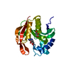
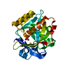
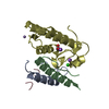
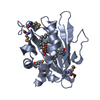
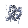
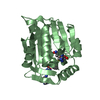
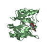
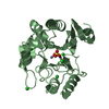
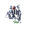
| Title | Crystal structure of the putative protein (Aq1419) from Aquifex aeolicus VF5 | ||||||
 Components Components | Probable molybdopterin-guanine dinucleotide biosynthesis protein A | ||||||
 Keywords Keywords | BIOSYNTHETIC PROTEIN / putative protein / Aq_1419 / molybdenum cofactor / Structural Genomics / NPPSFA / National Project on Protein Structural and Functional Analyses / RIKEN Structural Genomics/Proteomics Initiative / RSGI | ||||||
| Function / homology |  Function and homology information Function and homology informationmolybdenum cofactor guanylyltransferase / molybdenum cofactor guanylyltransferase activity / Mo-molybdopterin cofactor biosynthetic process / nucleotidyltransferase activity / GTP binding / metal ion binding / cytoplasm Similarity search - Function | ||||||
| Biological species |   Aquifex aeolicus (bacteria) Aquifex aeolicus (bacteria) | ||||||
| Method |  X-RAY DIFFRACTION / SYNCHROTRON / MAD / Resolution: 1.61 Å X-RAY DIFFRACTION / SYNCHROTRON / MAD / Resolution: 1.61 Å | ||||||
 Authors Authors | Kumarevel, T.S. / Malathy sony, S.M. / Ponnuswamy, M.N. / Kuramitsu, S. / Yokoyama, S. / RIKEN Structural Genomics/Proteomics Initiative (RSGI) | ||||||
 Citation Citation | Journal: To be Published Title: Crystal structure of the putative protein (Aq1419) from Aquifex aeolicus VF5 Authors: Kumarevel, T.S. / Malathy Sony, S.M. / Ponnuswamy, M.N. / Kuramitsu, S. / Yokoyama, S. | ||||||
| History |
|
- Structure visualization
| Structure viewer | Molecule: MolmilJmol/JSmol |
|---|
- Downloads & links
-Download
| PDBx/mmCIF format | 2e8b.cif.gz | 53.7 KB | Display | PDBx/mmCIF format |
|---|---|---|---|---|
| PDB format | pdb2e8b.ent.gz | 38.6 KB | Display | PDB format |
| PDBx/mmJSON format | 2e8b.json.gz | Tree view | PDBx/mmJSON format | |
| Others |  Other downloads Other downloads |
-Validation report
| Summary document | 2e8b_validation.pdf.gz | 429.1 KB | Display | wwPDB validaton report |
|---|---|---|---|---|
| Full document | 2e8b_full_validation.pdf.gz | 433.5 KB | Display | |
| Data in XML | 2e8b_validation.xml.gz | 11.8 KB | Display | |
| Data in CIF | 2e8b_validation.cif.gz | 16.5 KB | Display | |
| Arichive directory | https://data.pdbj.org/pub/pdb/validation_reports/e8/2e8bftp://data.pdbj.org/pub/pdb/validation_reports/e8/2e8b | HTTPS FTP |
-Related structure data
| Similar structure data | |
|---|---|
| Other databases |

-Links
 PDBj
PDBj

- Assembly
| Deposited unit | 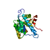
| ||||||||
|---|---|---|---|---|---|---|---|---|---|
| 1 |
| ||||||||
| Unit cell |
|
-Components
| #1: Protein | Mass: 23389.279 Da / Num. of mol.: 1 Source method: isolated from a genetically manipulated source Source: (gene. exp.) Aquifex aeolicus (bacteria) / Strain: VF5 / Gene: mobA / Plasmid: pET-21a / Production host: |
|---|---|
| #2: Water | ChemComp-HOH /  Mass: 18.015 Da / Num. of mol.: 192 / Source method: isolated from a natural source / Formula: H2O Mass: 18.015 Da / Num. of mol.: 192 / Source method: isolated from a natural source / Formula: H2O |
-Experimental details
-Experiment
| Experiment | Method: X-RAY DIFFRACTION / Number of used crystals: 1 |
|---|
- Sample preparation
| Crystal | Density Matthews: 1.95 Å3/Da / Density % sol: 36.77 % |
|---|---|
| Crystal grow | Temperature: 293 K / Method: vapor diffusion, sitting drop / pH: 6.2 Details: 0.2M Ammonium Iodide, 20% PEG3350, pH 6.2, VAPOR DIFFUSION, SITTING DROP, temperature 293K |
-Data collection
| Diffraction | Mean temperature: 100 K | ||||||||||||
|---|---|---|---|---|---|---|---|---|---|---|---|---|---|
| Diffraction source | Source: SYNCHROTRON / Site: SPring-8  / Beamline: BL26B2 / Wavelength: 0.97891, 0.97944, 0.9 / Beamline: BL26B2 / Wavelength: 0.97891, 0.97944, 0.9 | ||||||||||||
| Detector | Type: ADSC QUANTUM 210 / Detector: CCD / Date: Jun 14, 2006 | ||||||||||||
| Radiation | Monochromator: Si double crystal / Protocol: MAD / Monochromatic (M) / Laue (L): M / Scattering type: x-ray | ||||||||||||
| Radiation wavelength |
| ||||||||||||
| Reflection | Resolution: 1.61→42.1 Å / Num. all: 24264 / Num. obs: 24264 / % possible obs: 99.9 % / Observed criterion σ(F): 3 / Observed criterion σ(I): 3 / Redundancy: 6.82 % / Biso Wilson estimate: 21.3 Å2 / Rmerge(I) obs: 0.058 | ||||||||||||
| Reflection shell | Resolution: 1.61→1.67 Å / Redundancy: 6.23 % / Rmerge(I) obs: 0.341 / Num. unique all: 2403 / % possible all: 100 |
- Processing
| Software |
| ||||||||||||||||||||||||||||||||||||
|---|---|---|---|---|---|---|---|---|---|---|---|---|---|---|---|---|---|---|---|---|---|---|---|---|---|---|---|---|---|---|---|---|---|---|---|---|---|
| Refinement | Method to determine structure: MAD / Resolution: 1.61→39.62 Å / Rfactor Rfree error: 0.007 / Data cutoff high absF: 925066.14 / Data cutoff low absF: 0 / Isotropic thermal model: RESTRAINED / Cross valid method: THROUGHOUT / σ(F): 0 / Stereochemistry target values: Engh & Huber
| ||||||||||||||||||||||||||||||||||||
| Solvent computation | Solvent model: FLAT MODEL / Bsol: 46.6359 Å2 / ksol: 0.346911 e/Å3 | ||||||||||||||||||||||||||||||||||||
| Displacement parameters | Biso mean: 22.3 Å2
| ||||||||||||||||||||||||||||||||||||
| Refine analyze |
| ||||||||||||||||||||||||||||||||||||
| Refinement step | Cycle: LAST / Resolution: 1.61→39.62 Å
| ||||||||||||||||||||||||||||||||||||
| Refine LS restraints |
| ||||||||||||||||||||||||||||||||||||
| LS refinement shell | Resolution: 1.61→1.71 Å / Rfactor Rfree error: 0.021 / Total num. of bins used: 6
| ||||||||||||||||||||||||||||||||||||
| Xplor file |
|
