[English] 日本語
Yorodumi
- PDB-2dre: Crystal structure of Water-soluble chlorophyll protein from lepid... -

+
Open data


ID or keywords:

Loading...

-
Basic information

Entry
Database: PDB / ID: 2dre
TitleCrystal structure of Water-soluble chlorophyll protein from lepidium virginicum at 2.00 angstrom resolution
ComponentsWater-soluble chlorophyll protein
KeywordsPLANT PROTEIN / BETA-TREFOIL / TETRAMER / PLANT / LEPIDIUM VIRGINICUM / CHLOROPHYLL / WATER-SOLUBLE CHLOROPHYLL PROTEIN / SINGLET OXYGEN / PHOTOOXIDATION / CHLOROPHYLL CARRIER / KUNITZ (STI) INHIBITORS
Function / homology
Function and homology information


endopeptidase inhibitor activity
Similarity search - Function
Soybean trypsin inhibitor (Kunitz) protease inhibitors family signature. / Proteinase inhibitor I3, Kunitz legume / Trypsin and protease inhibitor / Soybean trypsin inhibitor (Kunitz) family of protease inhibitors / Kunitz inhibitor STI-like superfamily / Trefoil (Acidic Fibroblast Growth Factor, subunit A) - #50 / Trefoil (Acidic Fibroblast Growth Factor, subunit A) / Trefoil / Mainly Beta
Similarity search - Domain/homology
CHLOROPHYLL A / Water-soluble chlorophyll protein
Similarity search - Component
Biological speciesLepidium virginicum (plant)
MethodX-RAY DIFFRACTION / SYNCHROTRON / MIR / Resolution: 2 Å
AuthorsHorigome, D. / Satoh, H. / Itoh, N. / Mitsunaga, K. / Oonishi, I. / Nakagawa, A. / Uchida, A.
Citation
Journal: J.Biol.Chem. / Year: 2007
Title: Structural mechanism and photoprotective function of water-soluble chlorophyll-binding protein.
Authors: Horigome, D. / Satoh, H. / Itoh, N. / Mitsunaga, K. / Oonishi, I. / Nakagawa, A. / Uchida, A.
#1: Journal: Plant Cell.Physiol. / Year: 2001
Title: Water-soluble chlorophyll protein in Brassicaceae plants is a stress-induced chlorophyll-binding protein
Authors: Satoh, H. / Uchida, A. / Nakayama, K. / Okada, M.
#2: Journal: Biochim.Biophys.Acta / Year: 1980
Title: Crystallization of water-soluble chlorophyll-proteins from Lepidium virginicum
Authors: Murata, T. / Itoh, R. / Yakushiji, E.
History
DepositionJun 8, 2006Deposition site: PDBJ / Processing site: PDBJ
Revision 1.0Dec 26, 2006Provider: repository / Type: Initial release
Revision 1.1Apr 30, 2008Group: Version format compliance
Revision 1.2Jul 13, 2011Group: Version format compliance

-
Structure visualization

Structure viewerMolecule:
MolmilJmol/JSmol

Downloads & links

-
Assembly

Deposited unit
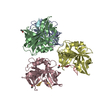
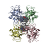
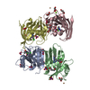
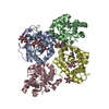
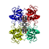
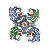
A: Water-soluble chlorophyll protein
B: Water-soluble chlorophyll protein
C: Water-soluble chlorophyll protein
D: Water-soluble chlorophyll protein
hetero molecules


Theoretical massNumber of molelcules
Total (without water)81,9408
Polymers78,3664
Non-polymers3,5744
Water9,998555
1


  • Idetical with deposited unit
  • defined by author&software
TypeNameSymmetry operationNumber
identity operation1_555x,y,z1
Buried area11900 Å2
ΔGint-98 kcal/mol
Surface area29830 Å2
MethodPISA
Unit cell
Length a, b, c (Å)73.060, 82.730, 121.880
Angle α, β, γ (deg.)90.00, 90.00, 90.00
Int Tables number19
Space group name H-MP212121

-
Components

#1: Protein
Water-soluble chlorophyll protein


Mass: 19591.527 Da / Num. of mol.: 4 / Fragment: RESIDUES 1-180 / Source method: isolated from a natural source / Source: (natural) Lepidium virginicum (plant) / Tissue: Leaf / References: UniProt: O04797
#2: Chemical
ChemComp-CLA / CHLOROPHYLL A


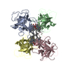
Mass: 893.489 Da / Num. of mol.: 4 / Source method: obtained synthetically / Formula: C55H72MgN4O5
#3: Water ChemComp-HOH / water


Mass: 18.015 Da / Num. of mol.: 555 / Source method: isolated from a natural source / Formula: H2O

-
Experimental details

-
Experiment

ExperimentMethod: X-RAY DIFFRACTION / Number of used crystals: 1

-
Sample preparation

CrystalDensity Matthews: 2.2 Å3/Da / Density % sol: 44.7 %
Crystal growTemperature: 293 K / Method: vapor diffusion, hanging drop / pH: 6
Details: 0.1M SODIUM/POTASSIUM PHOSPHATE, 3.2M AMMONIUM SULFATE, 5% SUCROSE, pH 6.00, VAPOR DIFFUSION, HANGING DROP, temperature 293K

-
Data collection

DiffractionMean temperature: 95 K
Diffraction sourceSource: SYNCHROTRON / Site: Photon Factory / Beamline: BL-6A / Wavelength: 0.978 / Wavelength: 0.978 Å
DetectorType: ADSC QUANTUM 4 / Detector: CCD / Date: Nov 6, 2003
RadiationMonochromator: TRIANGULAR SI(111) WITH AN ASYMMETRIC ANGLE OF 7.8 DEGREES
Protocol: SINGLE WAVELENGTH / Monochromatic (M) / Laue (L): M / Scattering type: x-ray
Radiation wavelengthWavelength: 0.978 Å / Relative weight: 1
ReflectionResolution: 2→28.99 Å / Num. obs: 50390 / % possible obs: 99.4 % / Observed criterion σ(I): -3 / Redundancy: 7 % / Biso Wilson estimate: 20.9 Å2 / Rmerge(I) obs: 0.068 / Rsym value: 0.063 / Net I/σ(I): 9.1
Reflection shellResolution: 2→2.11 Å / Redundancy: 6.8 % / Rmerge(I) obs: 0.533 / Mean I/σ(I) obs: 1.6 / Rsym value: 0.494 / % possible all: 99

-
Processing

Software
NameVersionClassification
HKL-2000data collection
SCALEPACKdata scaling
MLPHAREphasing
CNS1.1refinement
HKL-2000data reduction
RefinementMethod to determine structure: MIR / Resolution: 2→28.99 Å / Rfactor Rfree error: 0.006 / Data cutoff high absF: 1987876.77 / Data cutoff low absF: 0 / Isotropic thermal model: RESTRAINED / Cross valid method: THROUGHOUT / σ(F): 0 / Stereochemistry target values: ENGH & HUBER
Details: ALL OF THE FOUR CHLOROPHYLLS IN THE ASYMMETRIC UNIT WERE IDENTIFIED AS CHLOROPHYLL-A.
RfactorNum. reflection% reflectionSelection details
Rfree0.28 2489 4.9 %RANDOM
Rwork0.243 ---
obs0.243 50390 99.4 %-
Solvent computationSolvent model: FLAT MODEL / Bsol: 53.55 Å2 / ksol: 0.35 e/Å3
Displacement parametersBiso mean: 43.6 Å2
Baniso -1Baniso -2Baniso -3
1--3.31 Å20 Å20 Å2
2--1.22 Å20 Å2
3---2.1 Å2
Refine analyze
FreeObs
Luzzati coordinate error0.37 Å0.3 Å
Luzzati d res low-5 Å
Luzzati sigma a0.35 Å0.3 Å
Refinement stepCycle: LAST / Resolution: 2→28.99 Å
ProteinNucleic acidLigandSolventTotal
Num. atoms5353 0 260 555 6168
Refine LS restraints
Refine-IDTypeDev ideal
X-RAY DIFFRACTIONc_bond_d0.009
X-RAY DIFFRACTIONc_bond_d_na
X-RAY DIFFRACTIONc_bond_d_prot
X-RAY DIFFRACTIONc_angle_d
X-RAY DIFFRACTIONc_angle_d_na
X-RAY DIFFRACTIONc_angle_d_prot
X-RAY DIFFRACTIONc_angle_deg1.7
X-RAY DIFFRACTIONc_angle_deg_na
X-RAY DIFFRACTIONc_angle_deg_prot
X-RAY DIFFRACTIONc_dihedral_angle_d25
X-RAY DIFFRACTIONc_dihedral_angle_d_na
X-RAY DIFFRACTIONc_dihedral_angle_d_prot
X-RAY DIFFRACTIONc_improper_angle_d1.37
X-RAY DIFFRACTIONc_improper_angle_d_na
X-RAY DIFFRACTIONc_improper_angle_d_prot
X-RAY DIFFRACTIONc_mcbond_it
X-RAY DIFFRACTIONc_mcangle_it
X-RAY DIFFRACTIONc_scbond_it
X-RAY DIFFRACTIONc_scangle_it
LS refinement shellResolution: 2→2.13 Å / Rfactor Rfree error: 0.018 / Total num. of bins used: 6
RfactorNum. reflection% reflection
Rfree0.358 389 4.7 %
Rwork0.319 7825 -
obs--98.9 %
Xplor file
Refine-IDSerial noParam fileTopol file
X-RAY DIFFRACTION1PROTEIN_REP.PARAMPROTEIN.TOP
X-RAY DIFFRACTION2WATER_REP.PARAMWATER.TOP
X-RAY DIFFRACTION3ION.PARAMION.TOP
X-RAY DIFFRACTION4CLA_CNS_PARAMCLA_CNS_TOP

+
About Yorodumi

-
News

-
Feb 9, 2022. New format data for meta-information of EMDB entries

New format data for meta-information of EMDB entries

  • Version 3 of the EMDB header file is now the official format.
  • The previous official version 1.9 will be removed from the archive.

Related info.:EMDB header

External links:wwPDB to switch to version 3 of the EMDB data model

-
Aug 12, 2020. Covid-19 info

Covid-19 info

URL: https://pdbjlvh1.pdbj.org/emnavi/covid19.php

New page: Covid-19 featured information page in EM Navigator.

Related info.:Covid-19 info / Mar 5, 2020. Novel coronavirus structure data

+
Mar 5, 2020. Novel coronavirus structure data

Novel coronavirus structure data

Related info.:Yorodumi Speices / Aug 12, 2020. Covid-19 info

External links:COVID-19 featured content - PDBj / Molecule of the Month (242):Coronavirus Proteases

+
Jan 31, 2019. EMDB accession codes are about to change! (news from PDBe EMDB page)

EMDB accession codes are about to change! (news from PDBe EMDB page)

  • The allocation of 4 digits for EMDB accession codes will soon come to an end. Whilst these codes will remain in use, new EMDB accession codes will include an additional digit and will expand incrementally as the available range of codes is exhausted. The current 4-digit format prefixed with “EMD-” (i.e. EMD-XXXX) will advance to a 5-digit format (i.e. EMD-XXXXX), and so on. It is currently estimated that the 4-digit codes will be depleted around Spring 2019, at which point the 5-digit format will come into force.
  • The EM Navigator/Yorodumi systems omit the EMD- prefix.

Related info.:Q: What is EMD? / ID/Accession-code notation in Yorodumi/EM Navigator

External links:EMDB Accession Codes are Changing Soon! / Contact to PDBj

+
Jul 12, 2017. Major update of PDB

Major update of PDB

  • wwPDB released updated PDB data conforming to the new PDBx/mmCIF dictionary.
  • This is a major update changing the version number from 4 to 5, and with Remediation, in which all the entries are updated.
  • In this update, many items about electron microscopy experimental information are reorganized (e.g. em_software).
  • Now, EM Navigator and Yorodumi are based on the updated data.

External links:wwPDB Remediation / Enriched Model Files Conforming to OneDep Data Standards Now Available in the PDB FTP Archive

-
Yorodumi

Thousand views of thousand structures

  • Yorodumi is a browser for structure data from EMDB, PDB, SASBDB, etc.
  • This page is also the successor to EM Navigator detail page, and also detail information page/front-end page for Omokage search.
  • The word "yorodu" (or yorozu) is an old Japanese word meaning "ten thousand". "mi" (miru) is to see.

Related info.:EMDB / PDB / SASBDB / Comparison of 3 databanks / Yorodumi Search / Aug 31, 2016. New EM Navigator & Yorodumi / Yorodumi Papers / Jmol/JSmol / Function and homology information / Changes in new EM Navigator and Yorodumi

Read more