 Movie
Movie Controller

[English] 日本語
 Yorodumi
- PDB-2dqw: Crystal Structure of Dihydropteroate Synthase (FolP) from Thermus... -
+ Open data

- Basic information
| Entry | Database: PDB / ID: 2dqw | ||||||
|---|---|---|---|---|---|---|---|
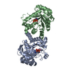
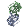
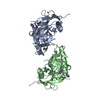
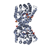
| Title | Crystal Structure of Dihydropteroate Synthase (FolP) from Thermus thermophilus HB8 | ||||||
 Components Components | Dihydropteroate synthase | ||||||
 Keywords Keywords | TRANSFERASE / Dihydropteroate Synthase / Dimer / Structural Genomics / NPPSFA / National Project on Protein Structural and Functional Analyses / RIKEN Structural Genomics/Proteomics Initiative / RSGI | ||||||
| Function / homology |  Function and homology information Function and homology informationdihydropteroate synthase / dihydropteroate synthase activity / folic acid biosynthetic process / tetrahydrofolate biosynthetic process / metal ion binding / cytosol Similarity search - Function | ||||||
| Biological species |   Thermus thermophilus (bacteria) Thermus thermophilus (bacteria) | ||||||
| Method |  X-RAY DIFFRACTION / SYNCHROTRON / MOLECULAR REPLACEMENT / Resolution: 1.65 Å X-RAY DIFFRACTION / SYNCHROTRON / MOLECULAR REPLACEMENT / Resolution: 1.65 Å | ||||||
 Authors Authors | Bagautdinov, B. / Kunishima, N. / RIKEN Structural Genomics/Proteomics Initiative (RSGI) | ||||||
 Citation Citation | Journal: To be Published Title: Crystal Structure of Dihydropteroate Synthase (FolP) from Thermus thermophilus HB8 Authors: Bagautdinov, B. / Kunishima, N. | ||||||
| History |
|
- Structure visualization
| Structure viewer | Molecule: MolmilJmol/JSmol |
|---|
- Downloads & links
-Download
| PDBx/mmCIF format | 2dqw.cif.gz | 120.7 KB | Display | PDBx/mmCIF format |
|---|---|---|---|---|
| PDB format | pdb2dqw.ent.gz | 92.6 KB | Display | PDB format |
| PDBx/mmJSON format | 2dqw.json.gz | Tree view | PDBx/mmJSON format | |
| Others |  Other downloads Other downloads |
-Validation report
| Arichive directory | https://data.pdbj.org/pub/pdb/validation_reports/dq/2dqwftp://data.pdbj.org/pub/pdb/validation_reports/dq/2dqw | HTTPS FTP |
|---|
-Related structure data
| Related structure data |  2dzaC  2dzbC  1eyeS S: Starting model for refinement C: citing same article ( |
|---|---|
| Similar structure data | |
| Other databases |


-Links
 PDBj
PDBj- Assembly
| Deposited unit | 
| ||||||||
|---|---|---|---|---|---|---|---|---|---|
| 1 |
| ||||||||
| Unit cell |
| ||||||||
| Details | The biological unit is a dimer and probably identical to the asymmetric unit |
-Components
| #1: Protein | Mass: 31897.020 Da / Num. of mol.: 2 Source method: isolated from a genetically manipulated source Source: (gene. exp.) Thermus thermophilus (bacteria) / Strain: HB8 / Gene: FolP / Plasmid: pET 11a / Species (production host): Escherichia coli / Production host: #2: Water | ChemComp-HOH / |  Mass: 18.015 Da / Num. of mol.: 611 / Source method: isolated from a natural source / Formula: H2O Mass: 18.015 Da / Num. of mol.: 611 / Source method: isolated from a natural source / Formula: H2O |
|---|
-Experimental details
-Experiment
| Experiment | Method: X-RAY DIFFRACTION / Number of used crystals: 1 |
|---|
- Sample preparation
| Crystal | Density Matthews: 2.42 Å3/Da / Density % sol: 49.09 % |
|---|---|
| Crystal grow | Temperature: 295 K / Method: microbatch / pH: 5.8 Details: PEG 20000, Mes, NaOH, pH 5.8, microbatch, temperature 295K |
-Data collection
| Diffraction | Mean temperature: 100 K |
|---|---|
| Diffraction source | Source: SYNCHROTRON / Site: SPring-8  / Beamline: BL26B1 / Wavelength: 1 Å / Beamline: BL26B1 / Wavelength: 1 Å |
| Detector | Type: MARRESEARCH / Detector: CCD / Date: May 21, 2006 / Details: mirrors |
| Radiation | Monochromator: GRAPHITE / Protocol: SINGLE WAVELENGTH / Monochromatic (M) / Laue (L): M / Scattering type: x-ray |
| Radiation wavelength | Wavelength: 1 Å / Relative weight: 1 |
| Reflection | Resolution: 1.65→40 Å / Num. all: 72924 / Num. obs: 72756 / % possible obs: 99.8 % / Observed criterion σ(F): 0 / Observed criterion σ(I): 0 / Redundancy: 9.5 % / Biso Wilson estimate: 25.5 Å2 / Rmerge(I) obs: 0.058 / Rsym value: 0.056 / Net I/σ(I): 17.7 |
| Reflection shell | Resolution: 1.65→1.71 Å / Redundancy: 8.9 % / Rmerge(I) obs: 0.494 / Mean I/σ(I) obs: 2.8 / Num. unique all: 7277 / Rsym value: 0.443 / % possible all: 100 |
- Processing
| Software |
| |||||||||||||||||||||||||
|---|---|---|---|---|---|---|---|---|---|---|---|---|---|---|---|---|---|---|---|---|---|---|---|---|---|---|
| Refinement | Method to determine structure: MOLECULAR REPLACEMENT Starting model: PDB ENTRY 1EYE Resolution: 1.65→29.88 Å / Isotropic thermal model: Overall / Cross valid method: THROUGHOUT / σ(F): 0 / σ(I): 0 / Stereochemistry target values: Engh & Huber
| |||||||||||||||||||||||||
| Displacement parameters | Biso mean: 34.6 Å2
| |||||||||||||||||||||||||
| Refine analyze |
| |||||||||||||||||||||||||
| Refinement step | Cycle: LAST / Resolution: 1.65→29.88 Å
| |||||||||||||||||||||||||
| Refine LS restraints |
| |||||||||||||||||||||||||
| LS refinement shell | Resolution: 1.65→1.71 Å / Rfactor Rfree error: 0.015
|
