 Movie
Movie Controller

[English] 日本語
 Yorodumi
- PDB-2dpd: Crystal structure of the Replication Termination Protein in compl... -
+ Open data

- Basic information
| Entry | Database: PDB / ID: 2dpd | ||||||
|---|---|---|---|---|---|---|---|
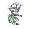
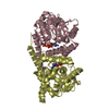
| Title | Crystal structure of the Replication Termination Protein in complex with a pseudosymmetric B-site | ||||||
 Components Components |
| ||||||
 Keywords Keywords | DNA BINDING PROTEIN/DNA / winged-helix protein-DNA complex / Replication Termination / Fork Arrest Mechanism / DNA BINDING PROTEIN-DNA COMPLEX | ||||||
| Function / homology |  Function and homology information Function and homology information | ||||||
| Biological species |  | ||||||
| Method |  X-RAY DIFFRACTION / SYNCHROTRON / MOLECULAR REPLACEMENT / Resolution: 3.17 Å X-RAY DIFFRACTION / SYNCHROTRON / MOLECULAR REPLACEMENT / Resolution: 3.17 Å | ||||||
 Authors Authors | Vivian, J.P. / Wilce, J. / Wilce, M.C.J. | ||||||
 Citation Citation | Journal: to be published Title: Crystal structure of the Replication Termination Protein in complex with a pseudosymmetric B-site Authors: Vivian, J.P. / Wilce, J. / Wilce, M.C.J. | ||||||
| History |
|
- Structure visualization
| Structure viewer | Molecule: MolmilJmol/JSmol |
|---|
- Downloads & links
-Download
| PDBx/mmCIF format | 2dpd.cif.gz | 157.7 KB | Display | PDBx/mmCIF format |
|---|---|---|---|---|
| PDB format | pdb2dpd.ent.gz | 123.9 KB | Display | PDB format |
| PDBx/mmJSON format | 2dpd.json.gz | Tree view | PDBx/mmJSON format | |
| Others |  Other downloads Other downloads |
-Validation report
| Arichive directory | https://data.pdbj.org/pub/pdb/validation_reports/dp/2dpdftp://data.pdbj.org/pub/pdb/validation_reports/dp/2dpd | HTTPS FTP |
|---|
-Related structure data
| Related structure data | 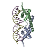 1f4kS S: Starting model for refinement |
|---|---|
| Similar structure data |


-Links
 PDBj
PDBj


- Assembly
| Deposited unit | 
| |||||||||||||||||||||||||||||||||||||||||||||||||||||||||||||||||
|---|---|---|---|---|---|---|---|---|---|---|---|---|---|---|---|---|---|---|---|---|---|---|---|---|---|---|---|---|---|---|---|---|---|---|---|---|---|---|---|---|---|---|---|---|---|---|---|---|---|---|---|---|---|---|---|---|---|---|---|---|---|---|---|---|---|---|
| 1 |
| |||||||||||||||||||||||||||||||||||||||||||||||||||||||||||||||||
| Unit cell |
| |||||||||||||||||||||||||||||||||||||||||||||||||||||||||||||||||
| Noncrystallographic symmetry (NCS) | NCS domain:
NCS domain segments: Ens-ID: 1 / Refine code: 1
| |||||||||||||||||||||||||||||||||||||||||||||||||||||||||||||||||
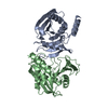
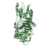
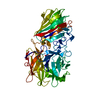
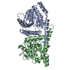
| Details | This structure represents one dimer of the functional termination complex which comprises a dimer of dimers |
-Components
| #1: DNA chain | Mass: 6445.209 Da / Num. of mol.: 1 / Source method: obtained synthetically | ||
|---|---|---|---|
| #2: DNA chain | Mass: 6436.195 Da / Num. of mol.: 1 / Source method: obtained synthetically | ||
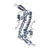
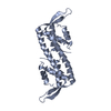
| #3: Protein | Mass: 14531.099 Da / Num. of mol.: 2 / Mutation: C110S Source method: isolated from a genetically manipulated source Source: (gene. exp.) #4: Water | ChemComp-HOH / |  Mass: 18.015 Da / Num. of mol.: 12 / Source method: isolated from a natural source / Formula: H2O Mass: 18.015 Da / Num. of mol.: 12 / Source method: isolated from a natural source / Formula: H2O |
-Experimental details
-Experiment
| Experiment | Method: X-RAY DIFFRACTION / Number of used crystals: 1 |
|---|
- Sample preparation
| Crystal | Density Matthews: 3.86 Å3/Da / Density % sol: 68.13 % | ||||||||||||||||||||||||||||||||||||
|---|---|---|---|---|---|---|---|---|---|---|---|---|---|---|---|---|---|---|---|---|---|---|---|---|---|---|---|---|---|---|---|---|---|---|---|---|---|
| Crystal grow | Temperature: 298 K / Method: vapor diffusion, hanging drop / pH: 4.6 Details: 25% MPD, 75mM sodium acetate pH 4.6, 20mM calcium chloride, VAPOR DIFFUSION, HANGING DROP, temperature 298K | ||||||||||||||||||||||||||||||||||||
| Components of the solutions |
|
-Data collection
| Diffraction | Mean temperature: 100 K |
|---|---|
| Diffraction source | Source: SYNCHROTRON / Site: APS  / Beamline: 14-BM-C / Wavelength: 1 Å / Beamline: 14-BM-C / Wavelength: 1 Å |
| Detector | Type: ADSC QUANTUM 4 / Detector: CCD / Date: Dec 5, 2001 |
| Radiation | Protocol: SINGLE WAVELENGTH / Monochromatic (M) / Laue (L): M / Scattering type: x-ray |
| Radiation wavelength | Wavelength: 1 Å / Relative weight: 1 |
| Reflection | Resolution: 3.15→60 Å / Num. all: 12241 / Num. obs: 11638 / % possible obs: 97.5 % / Observed criterion σ(F): 1 / Observed criterion σ(I): 1 / Redundancy: 6.5 % / Biso Wilson estimate: 57.1 Å2 / Rsym value: 0.069 / Net I/σ(I): 23.2 |
| Reflection shell | Resolution: 3.15→3.25 Å / Mean I/σ(I) obs: 3.3 / Rsym value: 0.365 / % possible all: 91.6 |
- Processing
| Software |
| |||||||||||||||||||||||||||||||||||||||||||||||||||||||||||||||||
|---|---|---|---|---|---|---|---|---|---|---|---|---|---|---|---|---|---|---|---|---|---|---|---|---|---|---|---|---|---|---|---|---|---|---|---|---|---|---|---|---|---|---|---|---|---|---|---|---|---|---|---|---|---|---|---|---|---|---|---|---|---|---|---|---|---|---|
| Refinement | Method to determine structure: MOLECULAR REPLACEMENT Starting model: PDB entry 1F4K with residues 71 to 89 of each monomer omitted Resolution: 3.17→60 Å / Cor.coef. Fo:Fc: 0.904 / Cor.coef. Fo:Fc free: 0.89 / SU B: 49.763 / SU ML: 0.393 / Cross valid method: THROUGHOUT / σ(F): 1 / ESU R: 0.792 / ESU R Free: 0.455 / Stereochemistry target values: MAXIMUM LIKELIHOOD
| |||||||||||||||||||||||||||||||||||||||||||||||||||||||||||||||||
| Solvent computation | Ion probe radii: 0.8 Å / Shrinkage radii: 0.8 Å / VDW probe radii: 1.4 Å / Solvent model: MASK | |||||||||||||||||||||||||||||||||||||||||||||||||||||||||||||||||
| Displacement parameters | Biso mean: 96.207 Å2
| |||||||||||||||||||||||||||||||||||||||||||||||||||||||||||||||||
| Refinement step | Cycle: LAST / Resolution: 3.17→60 Å
| |||||||||||||||||||||||||||||||||||||||||||||||||||||||||||||||||
| Refine LS restraints |
| |||||||||||||||||||||||||||||||||||||||||||||||||||||||||||||||||
| Refine LS restraints NCS | Number: 823 / Type: tight positional / Rms dev position: 0.05 Å / Weight position: 0.05 | |||||||||||||||||||||||||||||||||||||||||||||||||||||||||||||||||
| LS refinement shell | Resolution: 3.166→3.248 Å / Total num. of bins used: 20
|
