- PDB-2dp4: Crystal structure of the complex formed between proteinase K and ... -
+
Open data
ID or keywords:
Loading...
-
Basic information
Entry
Database: PDB / ID: 2dp4
Title
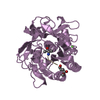
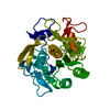
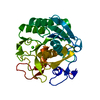
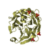
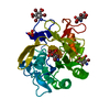
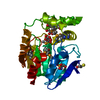
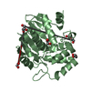
Crystal structure of the complex formed between proteinase K and a human lactoferrin fragment at 2.9 A resolution
Components
8-mer peptide from Lactotransferrin
Proteinase K
Keywords
HYDROLASE / proteinase K / lactoferrin fragment
Function / homology
Function and homology information
membrane destabilizing activity / host-mediated suppression of viral proces / Mtb iron assimilation by chelation / phagocytic vesicle lumen / Metal sequestration by antimicrobial proteins / negative regulation of viral process / negative regulation of tumor necrosis factor (ligand) superfamily member 11 production / positive regulation of toll-like receptor 4 signaling pathway / negative regulation of single-species biofilm formation in or on host organism / positive regulation of bone mineralization involved in bone maturation ...membrane destabilizing activity / host-mediated suppression of viral proces / Mtb iron assimilation by chelation / phagocytic vesicle lumen / Metal sequestration by antimicrobial proteins / negative regulation of viral process / negative regulation of tumor necrosis factor (ligand) superfamily member 11 production / positive regulation of toll-like receptor 4 signaling pathway / negative regulation of single-species biofilm formation in or on host organism / positive regulation of bone mineralization involved in bone maturation / negative regulation of ATP-dependent activity / negative regulation of osteoclast development / specific granule / negative regulation of lipopolysaccharide-mediated signaling pathway / positive regulation of chondrocyte proliferation / antifungal humoral response / regulation of tumor necrosis factor production / peptidase K / bone morphogenesis / Antimicrobial peptides / positive regulation of osteoblast proliferation / negative regulation of viral genome replication / positive regulation of protein serine/threonine kinase activity / humoral immune response / Hydrolases; Acting on peptide bonds (peptidases); Serine endopeptidases / positive regulation of osteoblast differentiation / cysteine-type endopeptidase inhibitor activity / : / regulation of cytokine production / secretory granule / ossification / protein serine/threonine kinase activator activity / innate immune response in mucosa / iron ion transport / lipopolysaccharide binding / recycling endosome / specific granule lumen / tertiary granule lumen / antimicrobial humoral immune response mediated by antimicrobial peptide / heparin binding / antibacterial humoral response / killing of cells of another organism / defense response to Gram-negative bacterium / early endosome / positive regulation of canonical NF-kappaB signal transduction / iron ion binding / Amyloid fiber formation / serine-type endopeptidase activity / Neutrophil degranulation / negative regulation of apoptotic process / cell surface / protein-containing complex / proteolysis / : / DNA binding / extracellular exosome / extracellular region / metal ion binding / nucleus / plasma membrane / cytoplasm Similarity search - Function
ProteinaseK / Tritirachium alkaline proteinase / Endopeptidase K
Mass: 28930.783 Da / Num. of mol.: 1 / Source method: isolated from a natural source / Source: (natural) Engyodontium album (fungus) / References: UniProt: P06873, peptidase K
#2: Protein/peptide
8-merpeptidefromLactotransferrin
Mass: 876.846 Da / Num. of mol.: 1 / Source method: isolated from a natural source Details: this peptiede was trapped durung its digestion by proteinase K. Source: (natural) Homo sapiens (human) / References: UniProt: P02788
In the structure databanks used in Yorodumi, some data are registered as the other names, "COVID-19 virus" and "2019-nCoV". Here are the details of the virus and the list of structure data.
Jan 31, 2019. EMDB accession codes are about to change! (news from PDBe EMDB page)
EMDB accession codes are about to change! (news from PDBe EMDB page)
The allocation of 4 digits for EMDB accession codes will soon come to an end. Whilst these codes will remain in use, new EMDB accession codes will include an additional digit and will expand incrementally as the available range of codes is exhausted. The current 4-digit format prefixed with “EMD-” (i.e. EMD-XXXX) will advance to a 5-digit format (i.e. EMD-XXXXX), and so on. It is currently estimated that the 4-digit codes will be depleted around Spring 2019, at which point the 5-digit format will come into force.
The EM Navigator/Yorodumi systems omit the EMD- prefix.
Related info.:Q: What is EMD? / ID/Accession-code notation in Yorodumi/EM Navigator
Yorodumi is a browser for structure data from EMDB, PDB, SASBDB, etc.
This page is also the successor to EM Navigator detail page, and also detail information page/front-end page for Omokage search.
The word "yorodu" (or yorozu) is an old Japanese word meaning "ten thousand". "mi" (miru) is to see.
Related info.:EMDB / PDB / SASBDB / Comparison of 3 databanks / Yorodumi Search / Aug 31, 2016. New EM Navigator & Yorodumi / Yorodumi Papers / Jmol/JSmol / Function and homology information / Changes in new EM Navigator and Yorodumi
 Movie
Movie Controller

 Yorodumi
 Open data
 Basic information
 Components
 Keywords
 Function and homology information
 Engyodontium album (fungus)
 Homo sapiens (human)
 X-RAY DIFFRACTION /
  Authors
 Citation
 Structure visualization
 Downloads & links
 Other downloads
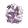


 PDBj
PDBj


 Assembly
 Mass: 18.015 Da / Num. of mol.: 112 / Source method: isolated from a natural source / Formula: H2O
 Sample preparation
 Processing