 Movie
Movie Controller

[English] 日本語
 Yorodumi
- PDB-2dhd: CRYSTALLOGRAPHIC ANALYSIS OF THE CATALYTIC MECHANISM OF HALOALKAN... -
+ Open data

- Basic information
| Entry | Database: PDB / ID: 2dhd | ||||||
|---|---|---|---|---|---|---|---|
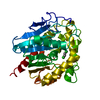
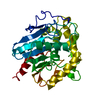
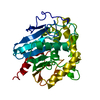
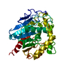
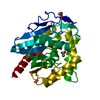
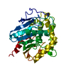
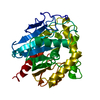
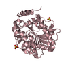
| Title | CRYSTALLOGRAPHIC ANALYSIS OF THE CATALYTIC MECHANISM OF HALOALKANE DEHALOGENASE | ||||||
 Components Components | HALOALKANE DEHALOGENASE | ||||||
 Keywords Keywords | DEHALOGENASE | ||||||
| Function / homology |  Function and homology information Function and homology information1,2-dichloroethane catabolic process / haloalkane dehalogenase / haloalkane dehalogenase activity / epoxide hydrolase activity / response to toxic substance Similarity search - Function | ||||||
| Biological species |  Xanthobacter autotrophicus (bacteria) Xanthobacter autotrophicus (bacteria) | ||||||
| Method |  X-RAY DIFFRACTION / Resolution: 2.13 Å X-RAY DIFFRACTION / Resolution: 2.13 Å | ||||||
 Authors Authors | Verschueren, K.H.G. / Dijkstra, B.W. | ||||||
 Citation Citation | Journal: Nature / Year: 1993 Title: Crystallographic analysis of the catalytic mechanism of haloalkane dehalogenase. Authors: Verschueren, K.H. / Seljee, F. / Rozeboom, H.J. / Kalk, K.H. / Dijkstra, B.W. #1: Journal: Embo J. / Year: 1991Title: Crystal Structure of Haloalkane Dehalogenase: An Enzyme to Detoxify Halogenated Alkanes Authors: Franken, S.M. / Rozeboom, H.J. / Kalk, K.H. / Dijkstra, B.W. #2: Journal: J.Mol.Biol. / Year: 1988Title: Crystallization of Haloalkane Dehalogenase from Xanthobacter Autotrophicus Gj10 Authors: Rozeboom, H.J. / Kingma, J. / Janssen, D.B. / Dijkstra, B.W. | ||||||
| History |
| ||||||
| Remark 700 | SHEET THE HELIX AND SHEET STRUCTURES ARE SIMILAR TO THOSE IN PROTEIN DATA BANK ENTRY 2HAD. |
- Structure visualization
| Structure viewer | Molecule: MolmilJmol/JSmol |
|---|
- Downloads & links
-Download
| PDBx/mmCIF format | 2dhd.cif.gz | 76.5 KB | Display | PDBx/mmCIF format |
|---|---|---|---|---|
| PDB format | pdb2dhd.ent.gz | 56.9 KB | Display | PDB format |
| PDBx/mmJSON format | 2dhd.json.gz | Tree view | PDBx/mmJSON format | |
| Others |  Other downloads Other downloads |
-Validation report
| Arichive directory | https://data.pdbj.org/pub/pdb/validation_reports/dh/2dhdftp://data.pdbj.org/pub/pdb/validation_reports/dh/2dhd | HTTPS FTP |
|---|
-Related structure data


-Links
 PDBj
PDBj


- Assembly
| Deposited unit | 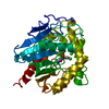
| ||||||||
|---|---|---|---|---|---|---|---|---|---|
| 1 |
| ||||||||
| Unit cell |
| ||||||||
| Atom site foot note | 1: CIS PROLINE - PRO 57 / 2: CIS PROLINE - PRO 168 |
-Components
| #1: Protein | Mass: 35238.297 Da / Num. of mol.: 1 Source method: isolated from a genetically manipulated source Source: (gene. exp.) Xanthobacter autotrophicus (bacteria) / References: UniProt: P22643, haloalkane dehalogenase |
|---|---|
| #2: Chemical | ChemComp-CL /   Mass: 35.453 Da / Num. of mol.: 1 / Source method: obtained synthetically / Formula: Cl Mass: 35.453 Da / Num. of mol.: 1 / Source method: obtained synthetically / Formula: Cl |
| #3: Water | ChemComp-HOH /  Mass: 18.015 Da / Num. of mol.: 151 / Source method: isolated from a natural source / Formula: H2O Mass: 18.015 Da / Num. of mol.: 151 / Source method: isolated from a natural source / Formula: H2O |
| Has protein modification | Y |
| Sequence details | SEQUENCE ADVISORY NOTICE: DIFFERENCE BETWEEN SWISS-PROT AND PDB SEQUENCE. SWISS-PROT ENTRY NAME: ...SEQUENCE ADVISORY NOTICE: DIFFERENCE |
-Experimental details
-Experiment
| Experiment | Method: X-RAY DIFFRACTION |
|---|
- Sample preparation
| Crystal | Density Matthews: 2.03 Å3/Da / Density % sol: 39.39 % | ||||||||||||||||||
|---|---|---|---|---|---|---|---|---|---|---|---|---|---|---|---|---|---|---|---|
| Crystal grow | *PLUS Temperature: 0 K / pH: 6.2 / Method: vapor diffusion, hanging drop | ||||||||||||||||||
| Components of the solutions | *PLUS
|
-Data collection
| Radiation | Scattering type: x-ray |
|---|---|
| Radiation wavelength | Relative weight: 1 |
| Reflection | *PLUS Highest resolution: 1.99 Å / Num. obs: 16812 / % possible obs: 82 % / Num. measured all: 39634 / Rmerge(I) obs: 0.0406 |
- Processing
| Software | Name: TNT / Classification: refinement | ||||||||||||||||||||||||||||||
|---|---|---|---|---|---|---|---|---|---|---|---|---|---|---|---|---|---|---|---|---|---|---|---|---|---|---|---|---|---|---|---|
| Refinement | Resolution: 2.13→15 Å / σ(F): 3 /
| ||||||||||||||||||||||||||||||
| Refinement step | Cycle: LAST / Resolution: 2.13→15 Å
| ||||||||||||||||||||||||||||||
| Refine LS restraints |
| ||||||||||||||||||||||||||||||
| Software | *PLUS Name: TNT / Classification: refinement | ||||||||||||||||||||||||||||||
| Refinement | *PLUS Rfactor obs: 0.189 | ||||||||||||||||||||||||||||||
| Solvent computation | *PLUS | ||||||||||||||||||||||||||||||
| Displacement parameters | *PLUS | ||||||||||||||||||||||||||||||
| Refine LS restraints | *PLUS Type: t_angle_d / Dev ideal: 3 |
