 Movie
Movie Controller

[English] 日本語
 Yorodumi
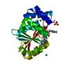
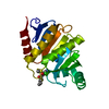
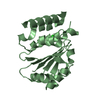
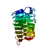
- PDB-2dbt: Crystal structure of chitinase C from Streptomyces griseus HUT6037 -
+ Open data

- Basic information
| Entry | Database: PDB / ID: 2dbt | ||||||
|---|---|---|---|---|---|---|---|
| Title | Crystal structure of chitinase C from Streptomyces griseus HUT6037 | ||||||
 Components Components | chitinase C | ||||||
 Keywords Keywords | HYDROLASE / family 19 chitinase | ||||||
| Function / homology |  Function and homology information Function and homology informationendochitinase activity / chitin catabolic process / defense response to fungus / cell wall macromolecule catabolic process / carbohydrate binding / carbohydrate metabolic process / extracellular region Similarity search - Function | ||||||
| Biological species |  Streptomyces griseus (bacteria) Streptomyces griseus (bacteria) | ||||||
| Method |  X-RAY DIFFRACTION / SYNCHROTRON / MOLECULAR REPLACEMENT / Resolution: 3.14 Å X-RAY DIFFRACTION / SYNCHROTRON / MOLECULAR REPLACEMENT / Resolution: 3.14 Å | ||||||
 Authors Authors | Kezuka, Y. / Watanabe, T. / Nonaka, T. | ||||||
 Citation Citation | Journal: J.Mol.Biol. / Year: 2006 Title: Structural Studies of a Two-domain Chitinase from Streptomyces griseus HUT6037 Authors: Kezuka, Y. / Ohishi, M. / Itoh, Y. / Watanabe, J. / Mitsutomi, M. / Watanabe, T. / Nonaka, T. #1: Journal: Biosci.Biotechnol.Biochem. / Year: 2002 Title: Functional analysis of the chitin-binding domain of a family 19 chitinase from Streptomyces griseus HUT6037: substrate-binding affinity and cis-dominant increase of antifungal function Authors: Itoh, Y. / Kawase, T. / Nikaidou, N. / Fukada, H. / Mitsutomi, M. / Watanabe, T. / Itoh, Y. #2: Journal: Microbiology / Year: 1999 Title: Family 19 chitinases of Streptomyces species: characterization and distribution Authors: Watanabe, T. / Kanai, R. / Kawase, T. / Tanabe, T. / Mitsutomi, M. / Sakuda, S. / Miyashita, K. #3: Journal: J.Bacteriol. / Year: 1996 Title: A modular family 19 chitinase found in the prokaryotic organism Streptomyces griseus HUT 6037 Authors: Ohno, T. / Armand, S. / Hata, T. / Nikaidou, N. / Henrissat, B. / Mitsutomi, M. / Watanabe, T. | ||||||
| History |
|
- Structure visualization
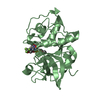
| Structure viewer | Molecule: MolmilJmol/JSmol |
|---|
- Downloads & links
-Download
| PDBx/mmCIF format | 2dbt.cif.gz | 126.9 KB | Display | PDBx/mmCIF format |
|---|---|---|---|---|
| PDB format | pdb2dbt.ent.gz | 99.9 KB | Display | PDB format |
| PDBx/mmJSON format | 2dbt.json.gz | Tree view | PDBx/mmJSON format | |
| Others |  Other downloads Other downloads |
-Validation report
| Summary document | 2dbt_validation.pdf.gz | 396.2 KB | Display | wwPDB validaton report |
|---|---|---|---|---|
| Full document | 2dbt_full_validation.pdf.gz | 406.2 KB | Display | |
| Data in XML | 2dbt_validation.xml.gz | 14.9 KB | Display | |
| Data in CIF | 2dbt_validation.cif.gz | 21.5 KB | Display | |
| Arichive directory | https://data.pdbj.org/pub/pdb/validation_reports/db/2dbtftp://data.pdbj.org/pub/pdb/validation_reports/db/2dbt | HTTPS FTP |
-Related structure data
| Related structure data |  1wvuC  1wvvSC C: citing same article ( S: Starting model for refinement |
|---|---|
| Similar structure data |


-Links
 PDBj
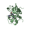
PDBj- Assembly
| Deposited unit | 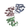
| ||||||||
|---|---|---|---|---|---|---|---|---|---|
| 1 | 
| ||||||||
| 2 | 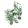
| ||||||||
| 3 | 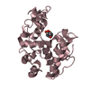
| ||||||||
| Unit cell |
|
-Components
| #1: Protein | Mass: 28497.170 Da / Num. of mol.: 3 / Fragment: residues 30-294 Source method: isolated from a genetically manipulated source Source: (gene. exp.) Streptomyces griseus (bacteria) / Plasmid: pET12a / Species (production host): Escherichia coli / Production host: #2: Chemical |   Mass: 238.305 Da / Num. of mol.: 3 / Source method: obtained synthetically / Formula: C8H18N2O4S / Comment: pH buffer*YM Mass: 238.305 Da / Num. of mol.: 3 / Source method: obtained synthetically / Formula: C8H18N2O4S / Comment: pH buffer*YM#3: Water | ChemComp-HOH / |  Mass: 18.015 Da / Num. of mol.: 21 / Source method: isolated from a natural source / Formula: H2O Mass: 18.015 Da / Num. of mol.: 21 / Source method: isolated from a natural source / Formula: H2OHas protein modification | Y | |
|---|
-Experimental details
-Experiment
| Experiment | Method: X-RAY DIFFRACTION / Number of used crystals: 1 |
|---|
- Sample preparation
| Crystal | Density Matthews: 3.56 Å3/Da / Density % sol: 65.47 % |
|---|---|
| Crystal grow | Temperature: 293 K / Method: vapor diffusion, hanging drop / pH: 7 Details: 2.3M Ammonium formate, 0.1M HEPES, pH 7.0, VAPOR DIFFUSION, HANGING DROP, temperature 293K |
-Data collection
| Diffraction | Mean temperature: 95 K |
|---|---|
| Diffraction source | Source: SYNCHROTRON / Site: Photon Factory  / Beamline: BL-6A / Wavelength: 0.978 Å / Beamline: BL-6A / Wavelength: 0.978 Å |
| Detector | Type: ADSC QUANTUM 4 / Detector: CCD / Date: Jan 27, 2002 |
| Radiation | Monochromator: Triangular Si(111) / Protocol: SINGLE WAVELENGTH / Monochromatic (M) / Laue (L): M / Scattering type: x-ray |
| Radiation wavelength | Wavelength: 0.978 Å / Relative weight: 1 |
| Reflection | Resolution: 3.14→58.32 Å / Num. all: 20454 / Num. obs: 20454 / % possible obs: 100 % / Observed criterion σ(F): 0 / Observed criterion σ(I): 0 / Redundancy: 10.5 % / Rmerge(I) obs: 0.074 / Rsym value: 0.074 / Net I/σ(I): 9.8 |
| Reflection shell | Resolution: 3.14→3.221 Å / Redundancy: 10.6 % / Rmerge(I) obs: 0.247 / Mean I/σ(I) obs: 3.1 / Num. unique all: 1497 / Rsym value: 0.247 / % possible all: 100 |
- Processing
| Software |
| ||||||||||||||||||||||||||||||||||||||||||||||||||||||||||||||||||||||||||||||||||||||||||
|---|---|---|---|---|---|---|---|---|---|---|---|---|---|---|---|---|---|---|---|---|---|---|---|---|---|---|---|---|---|---|---|---|---|---|---|---|---|---|---|---|---|---|---|---|---|---|---|---|---|---|---|---|---|---|---|---|---|---|---|---|---|---|---|---|---|---|---|---|---|---|---|---|---|---|---|---|---|---|---|---|---|---|---|---|---|---|---|---|---|---|---|
| Refinement | Method to determine structure: MOLECULAR REPLACEMENT Starting model: PDB ENTRY 1WVV Resolution: 3.14→58.32 Å / Cor.coef. Fo:Fc: 0.932 / Cor.coef. Fo:Fc free: 0.884 / SU B: 13.353 / SU ML: 0.23 / Cross valid method: THROUGHOUT / σ(F): 0 / σ(I): 0 / ESU R: 1.075 / ESU R Free: 0.348 / Stereochemistry target values: MAXIMUM LIKELIHOOD
| ||||||||||||||||||||||||||||||||||||||||||||||||||||||||||||||||||||||||||||||||||||||||||
| Solvent computation | Ion probe radii: 0.8 Å / Shrinkage radii: 0.8 Å / VDW probe radii: 1.2 Å / Solvent model: MASK | ||||||||||||||||||||||||||||||||||||||||||||||||||||||||||||||||||||||||||||||||||||||||||
| Displacement parameters | Biso mean: 41.688 Å2
| ||||||||||||||||||||||||||||||||||||||||||||||||||||||||||||||||||||||||||||||||||||||||||
| Refinement step | Cycle: LAST / Resolution: 3.14→58.32 Å
| ||||||||||||||||||||||||||||||||||||||||||||||||||||||||||||||||||||||||||||||||||||||||||
| Refine LS restraints |
| ||||||||||||||||||||||||||||||||||||||||||||||||||||||||||||||||||||||||||||||||||||||||||
| LS refinement shell | Resolution: 3.14→3.221 Å / Total num. of bins used: 20
|
