 Movie
Movie Controller

[English] 日本語
 Yorodumi
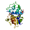
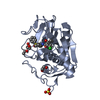
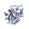
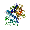
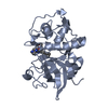
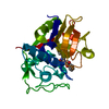
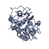
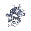
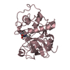
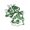
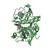
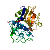
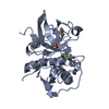
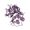
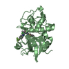
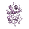
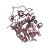
- PDB-4w5c: Crystal structure analysis of cruzain with three Fragments: 1 (N-... -
+ Open data

- Basic information
| Entry | Database: PDB / ID: 4w5c | ||||||
|---|---|---|---|---|---|---|---|
| Title | Crystal structure analysis of cruzain with three Fragments: 1 (N-(1H-benzimidazol-2-yl)-1,3-dimethyl-pyrazole-4-carboxamide), 6 (2-amino-4,6-difluorobenzothiazole) and 9 (N-(1H-benzimidazol-2-yl)-3-(4-fluorophenyl)-1H-pyrazole-4-carboxamide). | ||||||
 Components Components | Cruzipain | ||||||
 Keywords Keywords | HYDROLASE/HYDROLASE Inhibitor / Cysteine protease / cruzain / fragments-based drug discovery / mutagenesis / SPR / HYDROLASE-HYDROLASE Inhibitor complex | ||||||
| Function / homology |  Function and homology information Function and homology information | ||||||
| Biological species |  | ||||||
| Method |  X-RAY DIFFRACTION / SYNCHROTRON / MOLECULAR REPLACEMENT / Resolution: 3.27 Å X-RAY DIFFRACTION / SYNCHROTRON / MOLECULAR REPLACEMENT / Resolution: 3.27 Å | ||||||
 Authors Authors | Tochowicz, A. / McKerrow, J.H. / Craik, C.S. | ||||||
 Citation Citation | Journal: To Be Published Title: Applying Fragments Based- Drug Design to identify multiple binding modes on cysteine protease. Authors: Tochowicz, A. / Lee, G.M. / Arkin, M.R. / Neitz, J. / McKerrow, J. / Craik, C.S. | ||||||
| History |
|
- Structure visualization
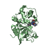
| Structure viewer | Molecule: MolmilJmol/JSmol |
|---|
- Downloads & links
-Download
| PDBx/mmCIF format | 4w5c.cif.gz | 410.8 KB | Display | PDBx/mmCIF format |
|---|---|---|---|---|
| PDB format | pdb4w5c.ent.gz | 342.2 KB | Display | PDB format |
| PDBx/mmJSON format | 4w5c.json.gz | Tree view | PDBx/mmJSON format | |
| Others |  Other downloads Other downloads |
-Validation report
| Arichive directory | https://data.pdbj.org/pub/pdb/validation_reports/w5/4w5cftp://data.pdbj.org/pub/pdb/validation_reports/w5/4w5c | HTTPS FTP |
|---|
-Related structure data
| Related structure data |  4w5bC  3kkuS S: Starting model for refinement C: citing same article ( |
|---|---|
| Similar structure data |


-Links
 PDBj
PDBj
- Assembly
| Deposited unit | 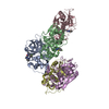
| ||||||||
|---|---|---|---|---|---|---|---|---|---|
| 1 | 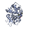
| ||||||||
| 2 | 
| ||||||||
| 3 | 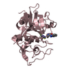
| ||||||||
| 4 | 
| ||||||||
| 5 | 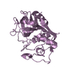
| ||||||||
| Unit cell |
|
-Components
| #1: Protein | Mass: 22756.121 Da / Num. of mol.: 5 / Fragment: UNP residues 122-337 / Mutation: C25S Source method: isolated from a genetically manipulated source Source: (gene. exp.)  #2: Chemical |   Mass: 255.275 Da / Num. of mol.: 2 / Source method: obtained synthetically / Formula: C13H13N5O Mass: 255.275 Da / Num. of mol.: 2 / Source method: obtained synthetically / Formula: C13H13N5O#3: Chemical | ChemComp-3H6 / | 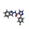  Mass: 321.308 Da / Num. of mol.: 1 / Source method: obtained synthetically / Formula: C17H12FN5O Mass: 321.308 Da / Num. of mol.: 1 / Source method: obtained synthetically / Formula: C17H12FN5O#4: Chemical |   Mass: 186.182 Da / Num. of mol.: 3 / Source method: obtained synthetically / Formula: C7H4F2N2S Mass: 186.182 Da / Num. of mol.: 3 / Source method: obtained synthetically / Formula: C7H4F2N2S#5: Water | ChemComp-HOH / |  Mass: 18.015 Da / Num. of mol.: 20 / Source method: isolated from a natural source / Formula: H2O Mass: 18.015 Da / Num. of mol.: 20 / Source method: isolated from a natural source / Formula: H2OHas protein modification | Y | |
|---|
-Experimental details
-Experiment
| Experiment | Method: X-RAY DIFFRACTION / Number of used crystals: 1 |
|---|
- Sample preparation
| Crystal | Density Matthews: 3.47 Å3/Da / Density % sol: 64.58 % |
|---|---|
| Crystal grow | Temperature: 293 K / Method: vapor diffusion, hanging drop / pH: 7.5 / Details: 0.1M Hepes pH 7.5, 1.2M K/Na Tartrate / PH range: 7.5 |
-Data collection
| Diffraction | Mean temperature: 100 K |
|---|---|
| Diffraction source | Source: SYNCHROTRON / Site: ALS  / Beamline: 8.3.1 / Wavelength: 1.115 Å / Beamline: 8.3.1 / Wavelength: 1.115 Å |
| Detector | Type: ADSC QUANTUM 315r / Detector: CCD / Date: Dec 8, 2012 |
| Radiation | Protocol: SINGLE WAVELENGTH / Monochromatic (M) / Laue (L): M / Scattering type: x-ray |
| Radiation wavelength | Wavelength: 1.115 Å / Relative weight: 1 |
| Reflection | Resolution: 3.27→30 Å / Num. obs: 25366 / % possible obs: 98.4 % / Redundancy: 8.1 % / Biso Wilson estimate: 84.44 Å2 / Rmerge(I) obs: 0.08 / Net I/σ(I): 17.8 |
| Reflection shell | Redundancy: 7.7 % / Mean I/σ(I) obs: 2.89 / % possible all: 99.1 |
- Processing
| Software |
| ||||||||||||||||||||||||||||||||||||||||||||||||||||||||
|---|---|---|---|---|---|---|---|---|---|---|---|---|---|---|---|---|---|---|---|---|---|---|---|---|---|---|---|---|---|---|---|---|---|---|---|---|---|---|---|---|---|---|---|---|---|---|---|---|---|---|---|---|---|---|---|---|---|
| Refinement | Method to determine structure: MOLECULAR REPLACEMENT Starting model: 3KKU Resolution: 3.27→29.98 Å / SU ML: 0.34 / Cross valid method: FREE R-VALUE / σ(F): 1.99 / Phase error: 27 / Stereochemistry target values: ML
| ||||||||||||||||||||||||||||||||||||||||||||||||||||||||
| Solvent computation | Shrinkage radii: 0.9 Å / VDW probe radii: 1.11 Å / Solvent model: FLAT BULK SOLVENT MODEL | ||||||||||||||||||||||||||||||||||||||||||||||||||||||||
| Displacement parameters | Biso mean: 86.96 Å2 | ||||||||||||||||||||||||||||||||||||||||||||||||||||||||
| Refinement step | Cycle: LAST / Resolution: 3.27→29.98 Å
| ||||||||||||||||||||||||||||||||||||||||||||||||||||||||
| Refine LS restraints |
| ||||||||||||||||||||||||||||||||||||||||||||||||||||||||
| LS refinement shell |
| ||||||||||||||||||||||||||||||||||||||||||||||||||||||||
| Refinement TLS params. | Method: refined / Origin x: -7.8522 Å / Origin y: 33.8031 Å / Origin z: 11.953 Å
| ||||||||||||||||||||||||||||||||||||||||||||||||||||||||
| Refinement TLS group | Selection details: ALL |
