Monochromator: Si double crystal / Protocol: SINGLE WAVELENGTH / Monochromatic (M) / Laue (L): M / Scattering type: x-ray
Radiation wavelength
Wavelength: 0.97892 Å / Relative weight: 1
Reflection
Redundancy: 7.4 % / Number: 84158 / Rmerge(I) obs: 0.08 / Χ2: 1.006 / D res high: 1.7 Å / D res low: 50 Å / % possible obs: 99.9
Diffraction reflection shell
Highest resolution (Å)
Lowest resolution (Å)
% possible obs (%)
ID
Rmerge(I) obs
Chi squared
Redundancy
3.66
50
99.9
1
0.053
0.999
7.2
2.91
3.66
100
1
0.06
0.999
7.5
2.54
2.91
100
1
0.075
1.002
7.5
2.31
2.54
99.9
1
0.088
1
7.5
2.14
2.31
99.9
1
0.101
1.002
7.5
2.02
2.14
100
1
0.125
1.006
7.5
1.91
2.02
100
1
0.171
1.006
7.5
1.83
1.91
100
1
0.238
1.011
7.5
1.76
1.83
100
1
0.344
1.012
7.4
1.7
1.76
99.8
1
0.432
1.023
6.5
Reflection
Resolution: 1.7→50 Å / Num. obs: 84158 / % possible obs: 99.9 % / Observed criterion σ(I): -3 / Redundancy: 7.4 % / Biso Wilson estimate: 15.8 Å2 / Rsym value: 0.08 / Χ2: 1.006 / Net I/σ(I): 21.94
Reflection shell
Resolution: 1.7→1.76 Å / Redundancy: 6.5 % / Mean I/σ(I) obs: 4.697 / Num. unique all: 8368 / Rsym value: 0.432 / Χ2: 1.023 / % possible all: 99.8
-
Processing
Software
Name
Version
Classification
NB
DENZO
datareduction
SCALEPACK
datascaling
CNS
1.1
refinement
PDB_EXTRACT
1.701
dataextraction
HKL-2000
datareduction
SOLVE
phasing
Refinement
Method to determine structure: SAD / Resolution: 1.7→36.29 Å / Data cutoff low absF: 0 / Cross valid method: THROUGHOUT / σ(F): 0 / Stereochemistry target values: Engh & Huber Details: THIS IS A TWINNED STRUCTURE. THE TWINNING OPERATOR IS (H,K,L) -> (H,-K,-L) AND THE TWINNING FRACTION IS 0.3201. THE R-FACTOR IS 0.1318 AND THE R-FREE IS 0.1431 WHEN THIS TWINNING OPERATOR IS USED.
In the structure databanks used in Yorodumi, some data are registered as the other names, "COVID-19 virus" and "2019-nCoV". Here are the details of the virus and the list of structure data.
Jan 31, 2019. EMDB accession codes are about to change! (news from PDBe EMDB page)
EMDB accession codes are about to change! (news from PDBe EMDB page)
The allocation of 4 digits for EMDB accession codes will soon come to an end. Whilst these codes will remain in use, new EMDB accession codes will include an additional digit and will expand incrementally as the available range of codes is exhausted. The current 4-digit format prefixed with “EMD-” (i.e. EMD-XXXX) will advance to a 5-digit format (i.e. EMD-XXXXX), and so on. It is currently estimated that the 4-digit codes will be depleted around Spring 2019, at which point the 5-digit format will come into force.
The EM Navigator/Yorodumi systems omit the EMD- prefix.
Related info.:Q: What is EMD? / ID/Accession-code notation in Yorodumi/EM Navigator
Yorodumi is a browser for structure data from EMDB, PDB, SASBDB, etc.
This page is also the successor to EM Navigator detail page, and also detail information page/front-end page for Omokage search.
The word "yorodu" (or yorozu) is an old Japanese word meaning "ten thousand". "mi" (miru) is to see.
Related info.:EMDB / PDB / SASBDB / Comparison of 3 databanks / Yorodumi Search / Aug 31, 2016. New EM Navigator & Yorodumi / Yorodumi Papers / Jmol/JSmol / Function and homology information / Changes in new EM Navigator and Yorodumi
 Movie
Movie Controller

 Yorodumi
 Open data
 Basic information
 Components
 Keywords
 Function and homology information
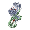
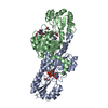
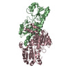
 Pyrococcus horikoshii (archaea)
 X-RAY DIFFRACTION /
  Authors
 Citation
 Structure visualization
 Downloads & links
 Other downloads
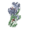


 PDBj
PDBj Assembly


 Mass: 96.063 Da / Num. of mol.: 2 / Source method: obtained synthetically / Formula: SO4
 Mass: 743.405 Da / Num. of mol.: 1 / Source method: obtained synthetically / Formula: C21H28N7O17P3
 Mass: 92.094 Da / Num. of mol.: 5 / Source method: obtained synthetically / Formula: C3H8O3
 Mass: 18.015 Da / Num. of mol.: 380 / Source method: isolated from a natural source / Formula: H2O
 Sample preparation
 / Beamline: BL26B1 / Wavelength: 0.97892 Å
 Processing