 Movie
Movie Controller

[English] 日本語
 Yorodumi
- PDB-2d7d: Structural insights into the cryptic DNA dependent ATP-ase activi... -
+ Open data

- Basic information
| Entry | Database: PDB / ID: 2d7d | ||||||
|---|---|---|---|---|---|---|---|
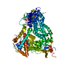
| Title | Structural insights into the cryptic DNA dependent ATP-ase activity of UvrB | ||||||
 Components Components |
| ||||||
 Keywords Keywords | HYDROLASE/DNA / Helicase / Protein-DNA-ADP ternary complex / HYDROLASE-DNA COMPLEX | ||||||
| Function / homology |  Function and homology information Function and homology informationnucleotide-excision repair, DNA damage recognition / excinuclease ABC activity / excinuclease repair complex / SOS response / ATP hydrolysis activity / DNA binding / ATP binding / cytoplasm Similarity search - Function | ||||||
| Biological species |  | ||||||
| Method |  X-RAY DIFFRACTION / SYNCHROTRON / MOLECULAR REPLACEMENT / Resolution: 2.1 Å X-RAY DIFFRACTION / SYNCHROTRON / MOLECULAR REPLACEMENT / Resolution: 2.1 Å | ||||||
 Authors Authors | Barrett, T.E. | ||||||
 Citation Citation | Journal: J.Mol.Biol. / Year: 2006 Title: Structural insights into the cryptic DNA-dependent ATPase activity of UvrB Authors: Eryilmaz, J. / Ceschini, S. / Ryan, J. / Geddes, S. / Waters, T.R. / Barrett, T.E. #1: Journal: To be PublishedTitle: Structural insights into the cryptic ATP-ase activity of UvrB Authors: Eryilmaz, J. / Ceschini, S. / Ryan, J. / Geddes, S. / Waters, T.R. / Barrett, T.E. | ||||||
| History |
|
- Structure visualization
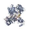
| Structure viewer | Molecule: MolmilJmol/JSmol |
|---|
- Downloads & links
-Download
| PDBx/mmCIF format | 2d7d.cif.gz | 149.9 KB | Display | PDBx/mmCIF format |
|---|---|---|---|---|
| PDB format | pdb2d7d.ent.gz | 114.3 KB | Display | PDB format |
| PDBx/mmJSON format | 2d7d.json.gz | Tree view | PDBx/mmJSON format | |
| Others |  Other downloads Other downloads |
-Validation report
| Arichive directory | https://data.pdbj.org/pub/pdb/validation_reports/d7/2d7dftp://data.pdbj.org/pub/pdb/validation_reports/d7/2d7d | HTTPS FTP |
|---|
-Related structure data
| Related structure data |  1d9xS S: Starting model for refinement |
|---|---|
| Similar structure data |


-Links
 PDBj
PDBj- Assembly
| Deposited unit | 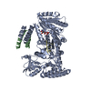
| ||||||||
|---|---|---|---|---|---|---|---|---|---|
| 1 |
| ||||||||
| Unit cell |
| ||||||||
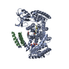
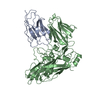
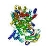
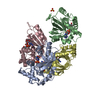
| Details | The biological assembly consists of a full UvrB monomer, a trithymine oligonucleotide, a single molecule of ADP and a helix-loop-helix dimer fragment resulting from proteolysis of UvrB. |
-Components
| #1: DNA chain | Mass: 867.621 Da / Num. of mol.: 1 / Source method: obtained synthetically |
|---|---|
| #2: Protein | Mass: 76440.133 Da / Num. of mol.: 1 Source method: isolated from a genetically manipulated source Source: (gene. exp.) References: UniProt: P37954, Hydrolases; Acting on ester bonds |
| #3: Protein/peptide | Mass: 4692.391 Da / Num. of mol.: 1 Source method: isolated from a genetically manipulated source Source: (gene. exp.) References: UniProt: P37954, Hydrolases; Acting on ester bonds |
| #4: Chemical | ChemComp-ADP /   Mass: 427.201 Da / Num. of mol.: 1 / Source method: obtained synthetically / Formula: C10H15N5O10P2 / Comment: ADP, energy-carrying molecule*YM Mass: 427.201 Da / Num. of mol.: 1 / Source method: obtained synthetically / Formula: C10H15N5O10P2 / Comment: ADP, energy-carrying molecule*YM |
| #5: Water | ChemComp-HOH /  Mass: 18.015 Da / Num. of mol.: 204 / Source method: isolated from a natural source / Formula: H2O Mass: 18.015 Da / Num. of mol.: 204 / Source method: isolated from a natural source / Formula: H2O |
-Experimental details
-Experiment
| Experiment | Method: X-RAY DIFFRACTION / Number of used crystals: 1 |
|---|
- Sample preparation
| Crystal | Density Matthews: 2.12 Å3/Da / Density % sol: 41.98 % | ||||||||||||||||||||||||||||
|---|---|---|---|---|---|---|---|---|---|---|---|---|---|---|---|---|---|---|---|---|---|---|---|---|---|---|---|---|---|
| Crystal grow | Temperature: 289 K / Method: microbatch / pH: 8.5 Details: 18-20% (w/v) PEG 10000 0.1M Tris-Hcl, pH 8.5, Microbatch, temperature 289K | ||||||||||||||||||||||||||||
| Components of the solutions |
|
-Data collection
| Diffraction | Mean temperature: 100 K |
|---|---|
| Diffraction source | Source: SYNCHROTRON / Site: ESRF  / Beamline: BM14 / Wavelength: 0.976 Å / Beamline: BM14 / Wavelength: 0.976 Å |
| Detector | Type: MARRESEARCH / Detector: CCD / Date: Apr 22, 2005 / Details: Mirrors |
| Radiation | Monochromator: Si 111 channel-cut monochromator / Protocol: SINGLE WAVELENGTH / Monochromatic (M) / Laue (L): M / Scattering type: x-ray |
| Radiation wavelength | Wavelength: 0.976 Å / Relative weight: 1 |
| Reflection | Resolution: 2.1→30 Å / Num. all: 39593 / Num. obs: 39593 / % possible obs: 96.1 % / Observed criterion σ(F): 2.4 / Observed criterion σ(I): 2.4 / Redundancy: 5.8 % / Biso Wilson estimate: 25 Å2 / Rmerge(I) obs: 0.082 / Rsym value: 0.082 / Net I/σ(I): 5.8 |
| Reflection shell | Resolution: 2.1→2.21 Å / Redundancy: 5.8 % / Rmerge(I) obs: 0.31 / Mean I/σ(I) obs: 2.4 / Num. unique all: 5487 / Rsym value: 0.31 / % possible all: 92.9 |
- Processing
| Software |
| ||||||||||||||||||||||||||||||||||||||||||||||||||||||||||||||||||||||||||||||||||||||||||
|---|---|---|---|---|---|---|---|---|---|---|---|---|---|---|---|---|---|---|---|---|---|---|---|---|---|---|---|---|---|---|---|---|---|---|---|---|---|---|---|---|---|---|---|---|---|---|---|---|---|---|---|---|---|---|---|---|---|---|---|---|---|---|---|---|---|---|---|---|---|---|---|---|---|---|---|---|---|---|---|---|---|---|---|---|---|---|---|---|---|---|---|
| Refinement | Method to determine structure: MOLECULAR REPLACEMENT Starting model: PDB entry 1D9X Resolution: 2.1→30 Å / Cor.coef. Fo:Fc: 0.925 / Cor.coef. Fo:Fc free: 0.884 / SU B: 7.237 / SU ML: 0.186 / Isotropic thermal model: Isotropic / Cross valid method: THROUGHOUT / σ(F): 0 / ESU R: 0.292 / ESU R Free: 0.236 / Stereochemistry target values: MAXIMUM LIKELIHOOD / Details: HYDROGENS HAVE BEEN ADDED IN THE RIDING POSITIONS
| ||||||||||||||||||||||||||||||||||||||||||||||||||||||||||||||||||||||||||||||||||||||||||
| Solvent computation | Ion probe radii: 0.8 Å / Shrinkage radii: 0.8 Å / VDW probe radii: 1.2 Å / Solvent model: MASK | ||||||||||||||||||||||||||||||||||||||||||||||||||||||||||||||||||||||||||||||||||||||||||
| Displacement parameters | Biso mean: 23.146 Å2
| ||||||||||||||||||||||||||||||||||||||||||||||||||||||||||||||||||||||||||||||||||||||||||
| Refine analyze |
| ||||||||||||||||||||||||||||||||||||||||||||||||||||||||||||||||||||||||||||||||||||||||||
| Refinement step | Cycle: LAST / Resolution: 2.1→30 Å
| ||||||||||||||||||||||||||||||||||||||||||||||||||||||||||||||||||||||||||||||||||||||||||
| Refine LS restraints |
| ||||||||||||||||||||||||||||||||||||||||||||||||||||||||||||||||||||||||||||||||||||||||||
| LS refinement shell | Resolution: 2.1→2.154 Å / Total num. of bins used: 20
|
